|
※サムネイル画像をクリックすると拡大画像が表示されます。
Figure 1 Western Blot Validation with Human Recombinant Protein
Loading: 5ng (Lane 1), 25ng (Lane 2) and 100ng (Lane 3) of human CD4 recombinant protein.Antibodies: CD4 PM-5201 (1 μg/mL), 1h incubation at RT in 5% NFDM/TBST.Secondary: Goat anti-mouse IgG HRP conjugate at 1:5000 dilution.Observed at around 27kD.

Figure 2 Western Blot Validation in Human Thymus Tissue Lysate
Loading: 15 μg of lysates per lane.Antibodies: CD4 PM-5201 (Lane 1: 0.5 μg/mL, Lane 2: 1 μg/mL and Lane 3: 2 μg/mL), 1h incubation at RT in 5% NFDM/TBST.Secondary: Goat anti-mouse IgG HRP conjugate at 1:5000 dilution.
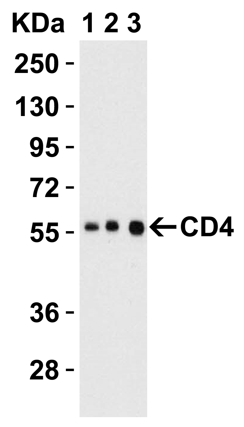
Figure 3 Western Blot Validation in Human Jurkat Lysate
Loading: 15 μg of lysates per lane.Antibodies: CD4 PM-5201 (Lane 1: 1 μg/mL, Lane 2: 2 μg/mL and Lane 3: 4 μg/mL), 1h incubation at RT in 5% NFDM/TBST.Secondary: Goat anti-mouse IgG HRP conjugate at 1:5000 dilution.

Figure 4 Western Blot Validation in Human, Mouse and Rat Tissue Lysate
Loading: 15 μg of lysates per lane.Antibodies: CD4 PM-5201 (1 μg/mL), 1h incubation at RT in 5% NFDM/TBST.Secondary: Goat anti-mouse IgG HRP conjugate at 1:5000 dilution.

Figure 5 Western Blot Validation in Human Cell Lines
Loading: 15 μg of lysates per lane.Antibodies: CD4 PM-5201 (4 μg/mL), 1h incubation at RT in 5% NFDM/TBST.Secondary: Goat anti-mouse IgG HRP conjugate at 1:5000 dilution.

Figure 6 Western Blot Validation in Mouse Spleen Tissue Lysate
Loading: 15 μg of lysates per lane.Antibodies: CD4 PM-5201 (Lane 1: 1 μg/mL, Lane 2: 2 μg/mL and Lane 3: 4 μg/mL), 1h incubation at RT in 5% NFDM/TBST.Secondary: Goat anti-mouse IgG HRP conjugate at 1:5000 dilution.

Figure 7 Western Blot Validation in Rat Lung Tissue Lysate
Loading: 15 μg of lysates per lane.Antibodies: CD4 PM-5201 (Lane 1: 0.5 μg/mL, Lane 2: 1 μg/mL and Lane 3: 2 μg/mL), 1h incubation at RT in 5% NFDM/TBST.Secondary: Goat anti-mouse IgG HRP conjugate at 1:5000 dilution.

Figure 8 Immunohistochemistry Validation of CD4 in Human Thymus Tissue
Immunohistochemical analysis of paraffin-embedded human thymus tissue using anti-CD4 antibody (PM-5201) at 5 μg/ml. Tissue was fixed with formaldehyde and blocked with 10% serum for 1 h at RT; antigen retrieval was by heat mediation with a citrate buffer (pH6). Samples were incubated with primary antibody overnight at 4℃. A goat anti-mouse IgG H&L (HRP) at 1/250 was used as secondary. Counter stained with Hematoxylin.

|

