|
※サムネイル画像をクリックすると拡大画像が表示されます。
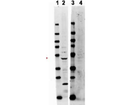
Western blot using Rockland's affinity purified anti-TRAF2 antibody shows detection of endogenous TRAF2 at ~47kDa (arrowhead). Lane 1 and 3: molecular weight markers. Lane 2: whole HeLa cell lysates (p/n W09-000-364). The identity of lower molecular weight band in lane 2 is unknown. Lane 4: incubated with immunizing peptide. Briefly, each lane contains approximately 14μg of lysate. Membranes were blocked in 3% BSA-TBS 30 min. at room temperature. Primary antibody was used at a 1:500 dilution in 3% BSA-TBS and reacted overnight at 4°C. The membrane was washed and reacted with a 1:20,000 dilution conjugated Gt-a-Rabbit DyLight 649 (p/n 611-143-122) for 1 hr at room temperature. Molecular weight estimation was made by comparison to prestained MW markers in lanes 1 and 3. Fluorescence image was captured using the VersaDocR Imaging System developed by Bio-Rad.
|
|
|
|
Western blot using Rockland's affinity purified anti-TRAF2 antibody shows detection of endogenous TRAF2 at ~47kDa (arrowhead). Lane 1 and 3: molecular weight markers. Lane 2: whole HeLa cell lysates (p/n W09-000-364). The identity of lower molecular weight band in lane 2 is unknown. Lane 4: incubated with immunizing peptide. Briefly, each lane contains approximately 14μg of lysate. Membranes were blocked in 3% BSA-TBS 30 min. at room temperature. Primary antibody was used at a 1:500 dilution in 3% BSA-TBS and reacted overnight at 4°C. The membrane was washed and reacted with a 1:20,000 dilution conjugated Gt-a-Rabbit DyLight 649 (p/n 611-143-122) for 1 hr at room temperature. Molecular weight estimation was made by comparison to prestained MW markers in lanes 1 and 3. Fluorescence image was captured using the VersaDocR Imaging System developed by Bio-Rad.
|
|
| 別品名 |
rabbit anti-TRAF2 antibody, E3 ubiquitin-protein ligase TRAF2, RING-type E3 ubiquitin transferase TRAF2, TNF receptor associated factor 2 antibody, TRAF-2, TRAF 2 antibody, TRAP-3, TRAP 3 antibody, TRAP3 antibody, Tumor necrosis factor type 2 receptor associated protein 3 antibody
|
| 交差種 |
Human
|
| 適用 |
Western Blot
Enzyme Linked Immunosorbent Assay
|
| 免疫動物 |
Rabbit
|
| 標識物 |
Unlabeled
|
| 精製度 |
Affinity Purified
|
| GENE ID |
7186
|
| Accession No.(Gene/Protein) |
22027612, Q12933
|
| Gene Symbol |
TRAF2
|
| 参考文献 |
Li S., Wang L., Berman M., Zhang Y., and Dorf M. (2006) RNAi Screen in Mouse Astrocytes Identifies Phosphatases that Regulate NF-κB Signaling. Mol Cell. Nov 17; 24:497-509.
|
| [注意事項] |
濃度はロットによって異なる可能性があります。メーカーDS及びCoAからご確認ください。
|
|
| メーカー |
品番 |
包装 |
|
RKL
|
600-401-B27S
|
25 UL
|
※表示価格について
| 当社在庫 |
なし
|
| 納期目安 |
約10日
|
| 保存温度 |
-20℃
|
|
※当社では商品情報の適切な管理に努めておりますが、表示される法規制情報は最新でない可能性があります。
また法規制情報の表示が無いものは、必ずしも法規制に非該当であることを示すものではありません。
商品のお届け前に最新の製品法規制情報をお求めの際はこちらへお問い合わせください。
|
※当社取り扱いの試薬・機器製品および受託サービス・創薬支援サービス(納品物、解析データ等)は、研究用としてのみ販売しております。
人や動物の医療用・臨床診断用・食品用としては、使用しないように、十分ご注意ください。
法規制欄に体外診断用医薬品と記載のものは除きます。
|
|
※リンク先での文献等のダウンロードに際しましては、掲載元の規約遵守をお願いします。
|
|
※CAS Registry Numbers have not been verified by CAS and may be inaccurate.
|
