|
※サムネイル画像をクリックすると拡大画像が表示されます。
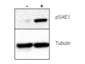
Western blot using Rockland's Rabbit anti-SAE1 pS185 antibody shows detection of phosphorylated SAE1. Left lane (-) contains 20 μg human HeLa whole cell protein. Right lane (+) contains 20 μg human HeLa whole cell protein from cells pre-treated with phosphatase inhibitor cocktail to prevent dephosphorylation of the target. Proteins were separated on a 10% SDS-PAGE and transferred onto nitrocellulose. After blocking with 5% milk-TBST 1 hr at room temperature, the membrane was probed with the primary antibody diluted to 1:1,000 at room temperature for 3 hr followed by washes and reaction with HRP-conjugated secondary and ECL imaging. Personal communication, Xin-Hua Feng, Baylor College of Medicine, Houston, TX
|
|
|
|
Western blot using Rockland's Rabbit anti-SAE1 pS185 antibody shows detection of phosphorylated SAE1. Left lane (-) contains 20 μg human HeLa whole cell protein. Right lane (+) contains 20 μg human HeLa whole cell protein from cells pre-treated with phosphatase inhibitor cocktail to prevent dephosphorylation of the target. Proteins were separated on a 10% SDS-PAGE and transferred onto nitrocellulose. After blocking with 5% milk-TBST 1 hr at room temperature, the membrane was probed with the primary antibody diluted to 1:1,000 at room temperature for 3 hr followed by washes and reaction with HRP-conjugated secondary and ECL imaging. Personal communication, Xin-Hua Feng, Baylor College of Medicine, Houston, TX
|
|
| 別品名 |
rabbit anti-SAE1 pS185 antibody, rabbit anti-SUMO activating enzyme subunit 1 pS185 antibody, Ubiquitin-like 1 activating enzyme E1A, UBLE1A, AOS1, SAE1, SUA1, SAE-1
|
| 交差種 |
Human
|
| 適用 |
Western Blot
Enzyme Linked Immunosorbent Assay
|
| 免疫動物 |
Rabbit
|
| 標識物 |
Unlabeled
|
| 精製度 |
Affinity Purified
|
| 翻訳後修飾 |
リン酸化
|
| GENE ID |
10055
|
| Accession No.(Gene/Protein) |
NP_005491.1, Q9UBE0
|
| Gene Symbol |
SAE1
|
| 参考文献 |
Wrighton KH,and Feng XH. (2006). Uba2. AfCS-Nature Molecule Pages. doi:10.1038/mp.a003681.01 Lois,L.M. and Lima,C.D. (2005) Structures of the SUMO E1 provide mechanistic insights into SUMO activation and E2 recruitment to E1. EMBO J. 24 (3), 439-451 Desterro,J.M., Rodriguez,M.S., Kemp,G.D. and Hay,R.T. (1999) Identification of the enzyme required for activation of the small ubiquitin-like protein SUMO-1. J. Biol. Chem. 274 (15), 10618-10624. Gong,L., Li,B., Millas,S. and Yeh,E.T. (1999) Molecular cloning and characterization of human AOS1 and UBA2, components of the sentrin-activating enzyme complex. FEBS Lett. 448 (1), 185-189.
|
|
| メーカー |
品番 |
包装 |
|
RKL
|
600-401-B24S
|
25 UL
|
※表示価格について
| 当社在庫 |
なし
|
| 納期目安 |
約10日
|
| 保存温度 |
-20℃
|
|
※当社では商品情報の適切な管理に努めておりますが、表示される法規制情報は最新でない可能性があります。
また法規制情報の表示が無いものは、必ずしも法規制に非該当であることを示すものではありません。
商品のお届け前に最新の製品法規制情報をお求めの際はこちらへお問い合わせください。
|
※当社取り扱いの試薬・機器製品および受託サービス・創薬支援サービス(納品物、解析データ等)は、研究用としてのみ販売しております。
人や動物の医療用・臨床診断用・食品用としては、使用しないように、十分ご注意ください。
法規制欄に体外診断用医薬品と記載のものは除きます。
|
|
※リンク先での文献等のダウンロードに際しましては、掲載元の規約遵守をお願いします。
|
|
※CAS Registry Numbers have not been verified by CAS and may be inaccurate.
|
