|
※サムネイル画像をクリックすると拡大画像が表示されます。
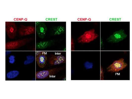
Immunofluorescence microscopy using Rockland's protein A purified anti-CENP-Q antibody shows detection of endogenous CENP-Q in HeLa whole cell lysate. Primary antibody was used at 1:100 followed by secondary antibody diluted 1:150. Red punctate anti-CENP-Q signal colocalizes in overlay images with green punctate anti-CREST signals at the kinetochores (attached points of sister chromatids). Visible are colocalized CENP-Q and CREST signal at various stages of the cell cycle as indicated from interphase to the end of mitosis. Nuclei are counter stained with bisbenzimide. Personal Communication, Kyung S. Lee, CCR-NCI, Bethesda, MD
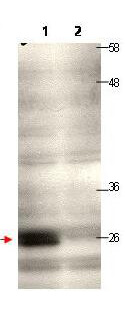
Western blot using Rockland's protein A purified anti-CENP-Q antibody shows detection of endogenous CENP-Q in a HeLa whole cell lysate (lane 1, arrowhead). The blot was incubated for 1.5 hours at room temperature using the primary antibody diluted to 0.5μg/mL, followed by washes and incubation with to the secondary antibody. Lane 1: Lysates from HeLa cells transfected with control sh-virus wherein the expression of CENP-Q is expected to not to alter. Lane 2: Lysates from HeLa cells transfected with Cenp-Q sh-virus wherein the expression of CENP-Q is knocked down significantly to a level where it is not being detected at all under the tested condition/WB exposure time. Personal Communication, Kyung S. Lee, CCR-NCI, Bethesda, MD.
|
|
|
|
Immunofluorescence microscopy using Rockland's protein A purified anti-CENP-Q antibody shows detection of endogenous CENP-Q in HeLa whole cell lysate. Primary antibody was used at 1:100 followed by secondary antibody diluted 1:150. Red punctate anti-CENP-Q signal colocalizes in overlay images with green punctate anti-CREST signals at the kinetochores (attached points of sister chromatids). Visible are colocalized CENP-Q and CREST signal at various stages of the cell cycle as indicated from interphase to the end of mitosis. Nuclei are counter stained with bisbenzimide. Personal Communication, Kyung S. Lee, CCR-NCI, Bethesda, MD
|
|
| 別品名 |
rabbit anti-CENP-Q Antibody, CenpQ, centromere protein Q, CENP Q
|
| 交差種 |
Human
|
| 適用 |
Western Blot
Enzyme Linked Immunosorbent Assay
Immuno Fluorescence
|
| 免疫動物 |
Rabbit
|
| 標識物 |
Unlabeled
|
| 精製度 |
Affinity Purified
|
| GENE ID |
55166
|
| Accession No.(Gene/Protein) |
40068061, Q7L2Z9
|
| Gene Symbol |
CENPQ
|
| 参考文献 |
[Pub Med ID]21454580
|
| [注意事項] |
濃度はロットによって異なる可能性があります。メーカーDS及びCoAからご確認ください。
|
|
| メーカー |
品番 |
包装 |
|
RKL
|
200-401-B48
|
100 UG
|
※表示価格について
| 当社在庫 |
なし
|
| 納期目安 |
約10日
|
| 保存温度 |
-20℃
|
|
※当社では商品情報の適切な管理に努めておりますが、表示される法規制情報は最新でない可能性があります。
また法規制情報の表示が無いものは、必ずしも法規制に非該当であることを示すものではありません。
商品のお届け前に最新の製品法規制情報をお求めの際はこちらへお問い合わせください。
|
※当社取り扱いの試薬・機器製品および受託サービス・創薬支援サービス(納品物、解析データ等)は、研究用としてのみ販売しております。
人や動物の医療用・臨床診断用・食品用としては、使用しないように、十分ご注意ください。
法規制欄に体外診断用医薬品と記載のものは除きます。
|
|
※リンク先での文献等のダウンロードに際しましては、掲載元の規約遵守をお願いします。
|
|
※CAS Registry Numbers have not been verified by CAS and may be inaccurate.
|
