|
※サムネイル画像をクリックすると拡大画像が表示されます。
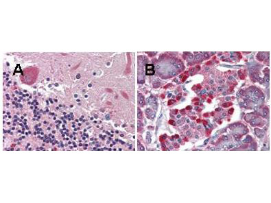
Rockland's anti-LGR4 monoclonal antibody was used diluted to 5 μg/ml to detect LGR4 staining at the membrane of cells in various human tissues. A. Brain cerebellum. B. Pancreas islet. Strongly positive staining is noted in subsets of cells within the islets of Langerhans. Moderately positive staining was observed in Purkinje and Golgi neurons of the cerebellum, adrenal medulla, neuroendocrine cells, hepatocytes, lung macrophages, seminiferous tubules and Leydig cells of the testis. Faintly to moderately positive staining was also observed in cardiac myocytes and renal tubules, granulocytes, and subsets of lymphocytes. Some elastin background staining is noted. Tissue was formalin fixed and paraffin embedded. No pre-treatment of sample was required. The image shows the localization of antibody as the precipitated red signal, with a hematoxylin purple nuclear counterstain. Personal communication, Andrew Elston, Lifespan Biosciences, Seattle, WA.
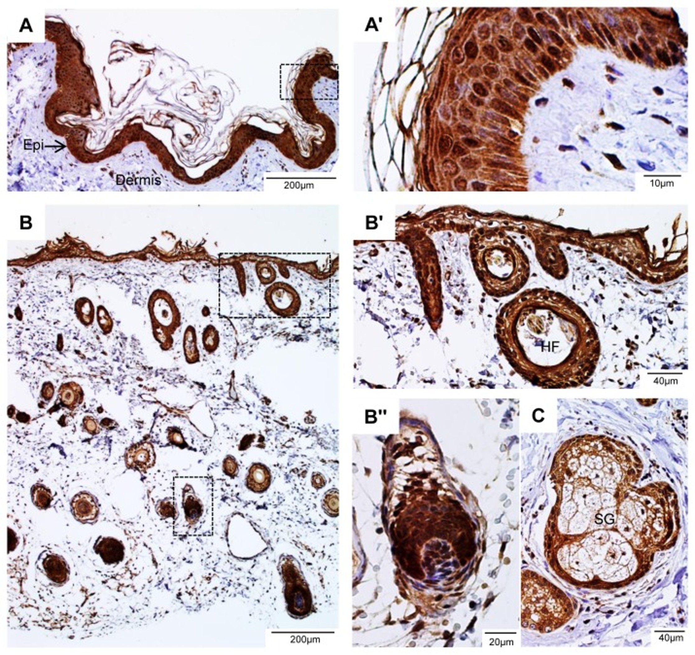
?LGR4 is highly expressed in the epidermis and hair follicles of human skin. A, representative micrograph of a skin section stained with 7E7, Epi ? epidermis. The boxed area is enlarged in A'. B, lower magnification of a skin section showing staining of hair follicles. The boxed areas are enlarged in B' and B'', HF ? hair follicle. C, staining was shown in the basal layer of sebaceous gland (SG).Fig 2. PMID:?24205130
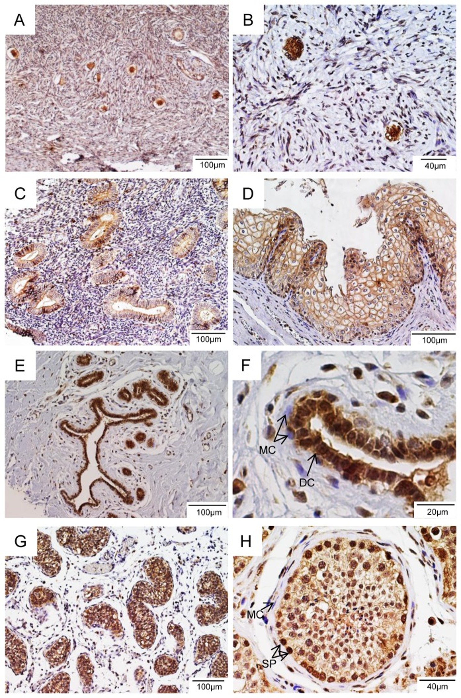
LGR4 is highly expressed in germ cells and epithelial cells in human reproductive organs. A, ovary, B, primordial follicles of ovary, C, uterine glands, D, stratified squamous epithelium of uterine cervix, E, mammary duct, F, breast intralobular terminal duct, DC ? duct cells, MC ? myoepithelial cells, G, testis, H, seminiferous tube of testis, SP ? spermatogonia, MC ? myoid cells. Fig 3. PMID:?24205130
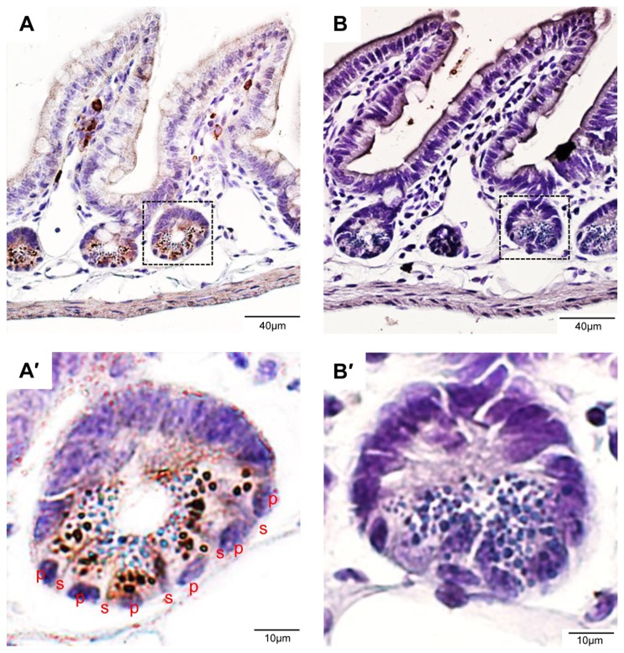
LGR4 is strongly expressed in the Paneth cells in mouse intestine. A, an ileum section stained with the antibody 5A3. B, an adjacent section stained with normal rat IgG. A' and B', enlarged view of the boxed area in A and B, respectively. P ? Paneth cells, s ? stem cells. Fig 4. PMID:?24205130
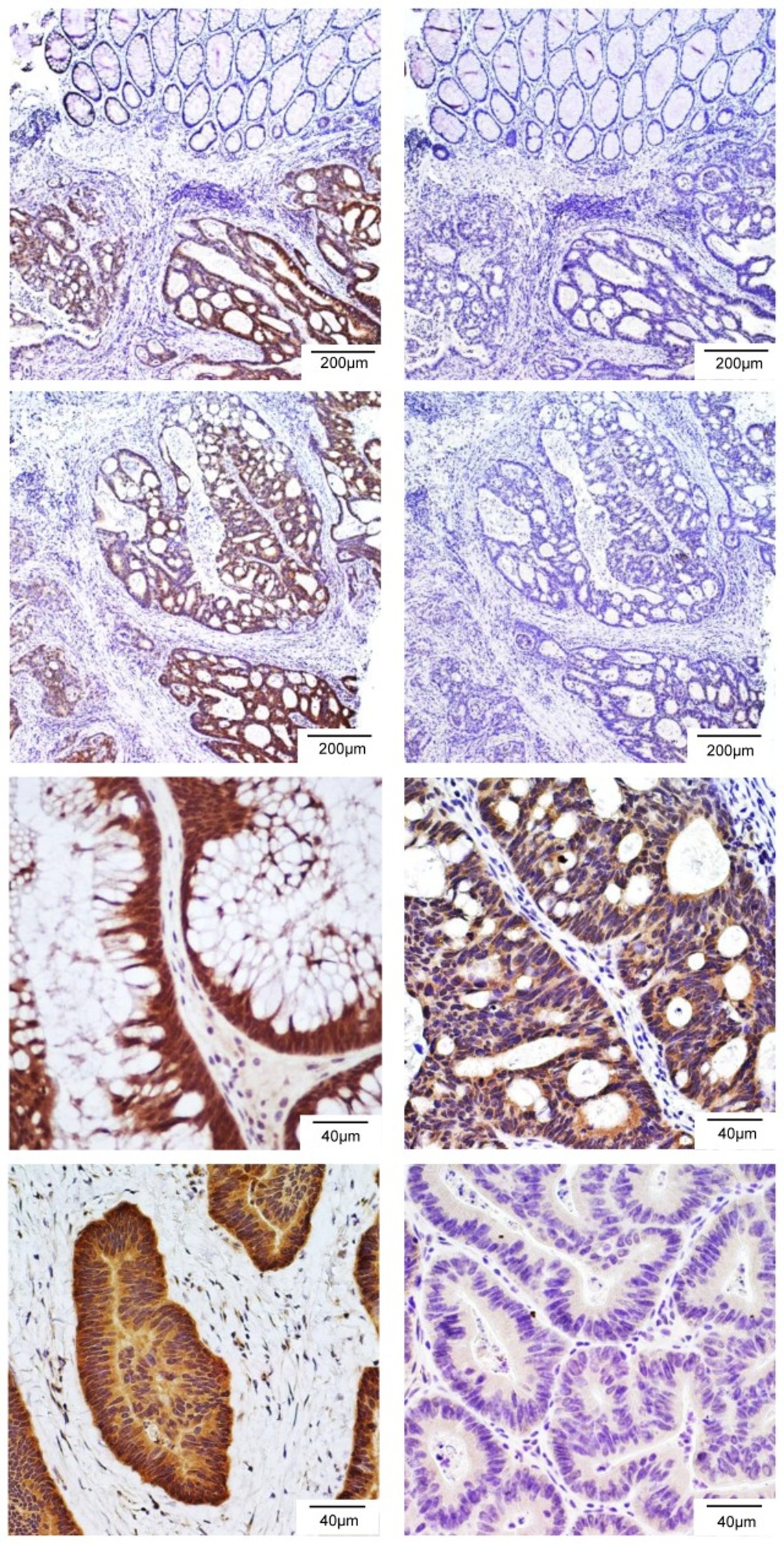
LGR4 is overexpressed in a significant fraction of human colon cancers. A and C, Staining of two colon tumor samples with 7E7. Specific and strong staining on colon tumor epithelial layers was clearly notable. B and D, Staining of adjacent sections of A and C, respectively, with 7E7 preincubated with human LGR4-ECD. The signal was blocked by incubation with LGR4-ECD. E-G, Three representatives of colon tumors with strong LGR4-IR. H, One representative of colon tumors with no expression of LGR4. Figure 5.PMID:?24205130
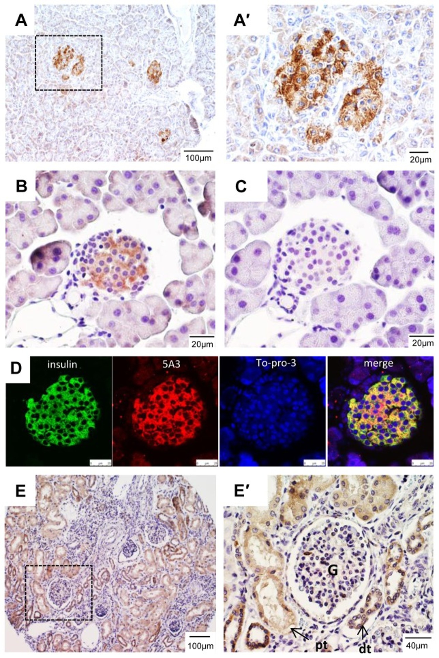
Expression of LGR4 in the pancreas and kidney. A, Staining of human pancreas with 7E7. A', Enlarged view of the area boxed in A. B, Staining of mouse pancreas with 5A3. C, Staining of an adjacent section of B with normal rat IgG as negative control. D, Co-staining of mouse pancreas with 5A3 and insulin antibody. E, Staining of human kidney, E', an enlarged view of the boxed area in E. G ? glomerulus, pt ? proximal convoluted tubule, dt ? distal convoluted tubule. Fig 6.PMID:?24205130
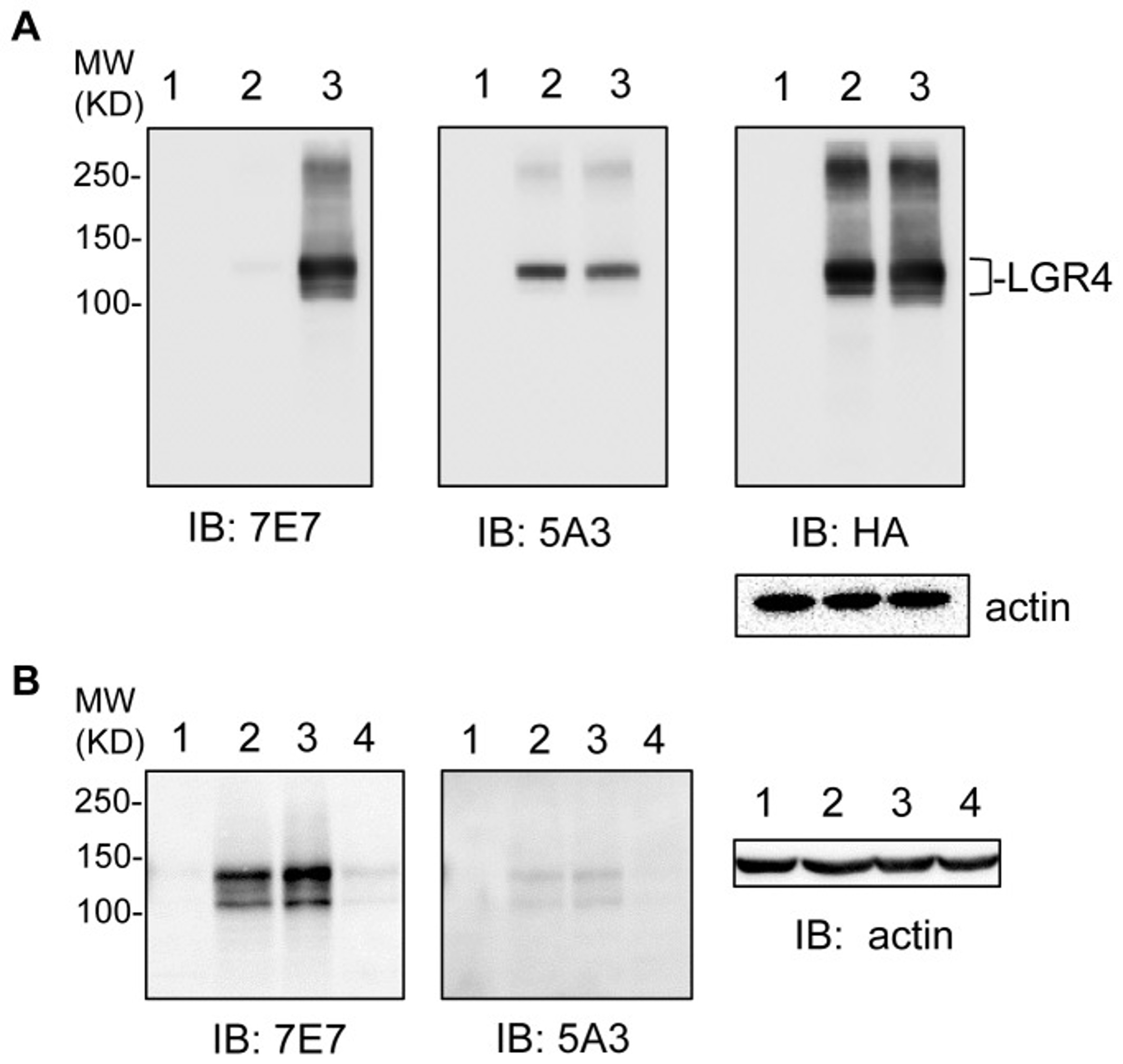
Recombinant and endogenous LGR4 were detected with custom generated rat monoclonal antibodies. A, Western blot of HEK293T transfected with vector (1), mouse LGR4 tagged with HA (2), or human LGR4 tagged with HA (3) using antibodies 7E7 (left panel), 5A3 (mid panel), and anti-HA (right panel). B, Western blot of LGR4 using 7E7 (left panel) and 5A3 (mid panel) on human colon tumor cell lines HCT116 (1), parental HT29 (2), vector-infected HT29 (3) and LGR4-shRNA infected HT29 (4). Actin was used as loading control. Fig 1. PMID:?24205130
|
|
|
|
Rockland's anti-LGR4 monoclonal antibody was used diluted to 5 μg/ml to detect LGR4 staining at the membrane of cells in various human tissues. A. Brain cerebellum. B. Pancreas islet. Strongly positive staining is noted in subsets of cells within the islets of Langerhans. Moderately positive staining was observed in Purkinje and Golgi neurons of the cerebellum, adrenal medulla, neuroendocrine cells, hepatocytes, lung macrophages, seminiferous tubules and Leydig cells of the testis. Faintly to moderately positive staining was also observed in cardiac myocytes and renal tubules, granulocytes, and subsets of lymphocytes. Some elastin background staining is noted. Tissue was formalin fixed and paraffin embedded. No pre-treatment of sample was required. The image shows the localization of antibody as the precipitated red signal, with a hematoxylin purple nuclear counterstain. Personal communication, Andrew Elston, Lifespan Biosciences, Seattle, WA.
|
|
| 別品名 |
mouse anti-LGR4 antibody, mouse anti-LGR 4 antibody, leucine-rich repeat-containing G protein-coupled receptor 4
|
| 別包装 |
あり
|
| 交差種 |
Human
|
| 適用 |
Western Blot
Enzyme Linked Immunosorbent Assay
|
| 免疫動物 |
Mouse
|
| クローン |
6G8.B3.G5.C3
|
| 抗体クラス |
IgG2bκ
|
| 標識物 |
Unlabeled
|
| 精製度 |
Ig fraction - Protein A
|
| GENE ID |
55366
|
| Accession No.(Gene/Protein) |
157694513, Q8N537
|
| Gene Symbol |
LGR4
|
| 参考文献 |
[[Cosmo Reference Collection]][Pub Med ID]24205130
|
| [注意事項] |
濃度はロットによって異なる可能性があります。メーカーDS及びCoAからご確認ください。
|
|
| メーカー |
品番 |
包装 |
|
RKL
|
200-301-B45S
|
25 UL
|
※表示価格について
| 当社在庫 |
なし
|
| 納期目安 |
約10日
|
| 保存温度 |
-20℃
|
|


