|
※サムネイル画像をクリックすると拡大画像が表示されます。
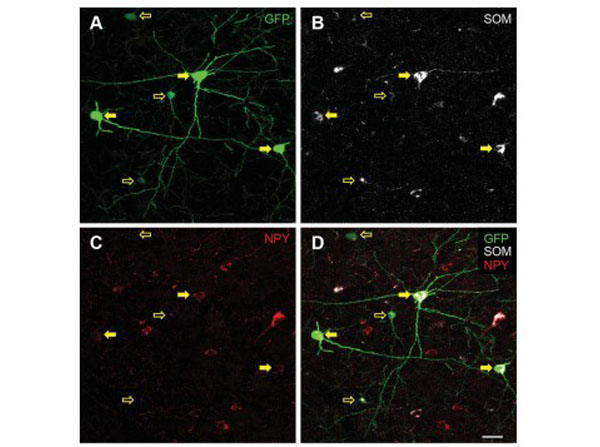
Analysis of neuropeptide Y (NPY) expressing interneurons in the cingulate cortex of FVB-Tg(GadGFP)45704Swn/J mice. A?D: Representative confocal images of cells immunopositive for GFP (green), somatostatin (SOM, white), and NPY (red), and merged image of all channels. Solid yellow arrows indicate GFP1/SOM1/NPY 1 cells and open yellow arrows GFP1/ SOM1/NPY2 cells. E: Mean 6 standard deviation of relative numbers of GFP1/SOM2/NPY2 cells, GFP1/SOM1/NPY2 cells, and GFP1/SOM1/NPY 1 cells in the cingulate cortex. Scale bar 5 20 lm in D (applies to A?D). Figure 10. PMID: 26669716.
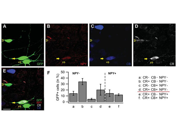
Analysis of coexpression of calretinin (CR), calbindin (CB), and NPY in GFP 1 cells in the cingulate cortex of FVBTg(GadGFP)45704Swn/J mice. A?E: Representative confocal images of cells in layers II?III of the cingulate cortex immunopositive for GFP (green), NPY (red), CR (white), and CB (blue). The solid yellow arrow indicates GFP1/CR1/CB1/NPY 1 cells, the open yellow arrow GFP1/CR2/CB2/NPY2 cells, and the open yellow arrowhead GFP1/CR1/CB1/NPY2 cells. F: Mean 6 standard deviation of relative numbers of GFP1/CR2/CB2/NPY2 cells (a), GFP1/CR1/CB2/NPY2 cells (b), GFP1/CR2/CB1/NPY2 cells (c), GFP1/CR1/ CB1/NPY2 cells (d), GFP1/CR1/CB2/NPY 1 cells (e), and GFP1/CR1/CB1/NPY 1 cells (f). Scale bar 5 20 lm in E (applies to A?E). Figure 11. PMID: 26669716.
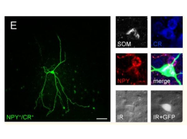
Morphological variety of group I GIN. A-E,?left panel?Confocal z-stack images as maximum intensity projections of representative group I GIN. Scalebars: 50 μm.?Right panel?Immunolabeling of biocytin-injected cells for GFP (green), CB (white or blue), CR (white or blue), SOM (white) or NPY (red). Fluorescence (white, GFP) and infrared (IR)-DIC (grey) images of recorded cells were acquired prior recording. Fig 8. PMID: 30001424.
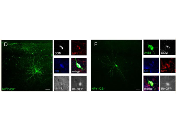
Morphological varieties in group II GIN. Many group I GIN classified as Martinotti cells with massive axonal arborizations in layer 1 and in the home layer. All scalebars: 50 μm.?A-F,?left panel?Confocal Z-stack images of biocytin-injected GIN as maximum intensity projections.?Right panel?Corresponding immunolabelings of the cells shown in the?left panel. Cells were labeled for GFP (green), CR (white or blue), CB (blue or white), SOM (white) or NPY (red). Fluorescence (GFP, white) and infrared-DIC (grey) images were acquired of cells in A-F prior recording. Fig 9. PMID: 30001424.
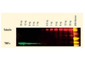
Dylight? dyes can be used for two-color Western Blot detection with low background and high signal. Anti-tubulin was detected using a DyLight? 549 conjugate. Anti-TNF? was detected using a DyLight? 649 conjugate. The image was captured using the Typhoon? 9410 Imaging System.
|
|
|
|
Analysis of neuropeptide Y (NPY) expressing interneurons in the cingulate cortex of FVB-Tg(GadGFP)45704Swn/J mice. A?D: Representative confocal images of cells immunopositive for GFP (green), somatostatin (SOM, white), and NPY (red), and merged image of all channels. Solid yellow arrows indicate GFP1/SOM1/NPY 1 cells and open yellow arrows GFP1/ SOM1/NPY2 cells. E: Mean 6 standard deviation of relative numbers of GFP1/SOM2/NPY2 cells, GFP1/SOM1/NPY2 cells, and GFP1/SOM1/NPY 1 cells in the cingulate cortex. Scale bar 5 20 lm in D (applies to A?D). Figure 10. PMID: 26669716.
|
|
| 別品名 |
Rabbit Anti-Sheep IgG DyLightTM 549 Conjugated Antibody, Rabbit Anti-Sheep IgG Antibody DyLightTM549 Conjugation
|
| 交差種 |
Sheep
|
| 免疫動物 |
Rabbit
|
| 標識物 |
DyLightTM 549
|
| 精製度 |
Affinity Purified
|
| 参考文献 |
[Pub Med ID]30001424
|
| [注意事項] |
濃度はロットによって異なる可能性があります。メーカーDS及びCoAからご確認ください。
|
|
| メーカー |
品番 |
包装 |
|
RKL
|
613-442-002
|
100 UG
|
※表示価格について
| 当社在庫 |
なし
|
| 納期目安 |
約10日
|
| 法規制 |
毒
|
| 保存温度 |
4℃
|
|
※当社では商品情報の適切な管理に努めておりますが、表示される法規制情報は最新でない可能性があります。
また法規制情報の表示が無いものは、必ずしも法規制に非該当であることを示すものではありません。
商品のお届け前に最新の製品法規制情報をお求めの際はこちらへお問い合わせください。
|
※当社取り扱いの試薬・機器製品および受託サービス・創薬支援サービス(納品物、解析データ等)は、研究用としてのみ販売しております。
人や動物の医療用・臨床診断用・食品用としては、使用しないように、十分ご注意ください。
法規制欄に体外診断用医薬品と記載のものは除きます。
|
|
※リンク先での文献等のダウンロードに際しましては、掲載元の規約遵守をお願いします。
|
|
※CAS Registry Numbers have not been verified by CAS and may be inaccurate.
|
