|
※サムネイル画像をクリックすると拡大画像が表示されます。
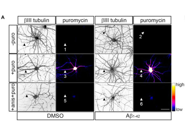
Detection of newly synthesized proteins by puromycilation.?(A)?Rat hippocampal neurons were grown for 9 DIV and were treated with DMSO (left panels) or Aβ1?42?oligomers (right panels) for 24 h. Before fixing, cells were incubated with vehicle (-puro; neurites 1 and 2), with puromycin (+ puro; neurites 3 and 4) or with puromycin and anisomycin (+ anis + puro; neurites 5 and 6) for 30 mins. Cells were immunostained with rabbit anti-βIII tubulin antibody (1:500) to visualize the neuronal cytoskeleton (gray) and with a mouse anti-puromycin antibody (1:500) to analyze newly synthesized proteins (heatmaps). Secondary anti-rabbit DyLight 405 (1:200, 611-146-002). Scale bar, 50 μm. Fig 2. PMID: 32581689.
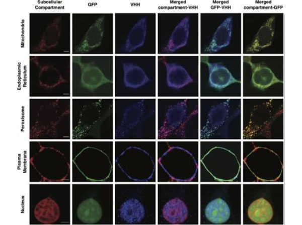
NanoLoc-mediated control of GFP subcellular localization. Representative confocal microscopy images of HEK293/GFP#1 cells transiently transfected for the expression of VHHLoc?variants and analyzed 72?h post-transfection. Subcellular compartment (red, column 1); GFP (green, column 2); VHH (blue, anti-HA, column 3); colocalization of subcellular compartment and VHH shown in merged images (purple, column 4); colocalization of GFP and VHH shown in merged images (cyan, column 5); colocalization of subcellular compartment and GFP shown in merged images (yellow, column 6). Scale bars: 5?μm. Brightness and contrast levels were adjusted and images of cells treated the same were subjected to the same adjustment. Pseudo-coloring was applied to the subcellular compartment stain and VHH images for the plasma membrane and the nucleus. Primary antibodies: rabbit anti-HA (VHH), 1:250, mouse anti-calnexin (endoplasmic reticulum membrane) 1:50; mouse anti-PMP70 (peroxisome) 1:50, with secondary antibodies: goat anti-rabbit DyLight 405 conjugated, (p/n 611-146-002) 1:200, goat anti-rabbit DyLight 549 conjugated, (p/n 611-142-002) 1:500, and goat anti-mouse DyLight 549 conjugated, (1:500). Fig 1. PMID: 33763602.
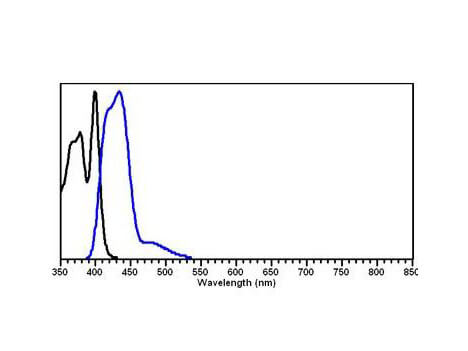
DyLight? 405 Fluorescence absorption
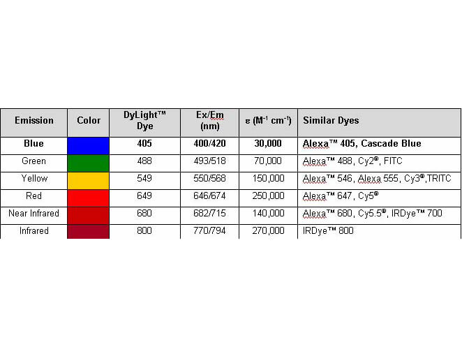
Properties of DyLight? Fluorescent Dyes.
|
|
|
|
Detection of newly synthesized proteins by puromycilation.?(A)?Rat hippocampal neurons were grown for 9 DIV and were treated with DMSO (left panels) or Aβ1?42?oligomers (right panels) for 24 h. Before fixing, cells were incubated with vehicle (-puro; neurites 1 and 2), with puromycin (+ puro; neurites 3 and 4) or with puromycin and anisomycin (+ anis + puro; neurites 5 and 6) for 30 mins. Cells were immunostained with rabbit anti-βIII tubulin antibody (1:500) to visualize the neuronal cytoskeleton (gray) and with a mouse anti-puromycin antibody (1:500) to analyze newly synthesized proteins (heatmaps). Secondary anti-rabbit DyLight 405 (1:200, 611-146-002). Scale bar, 50 μm. Fig 2. PMID: 32581689.
|
|
| 別品名 |
Goat anti-Rabbit IgG Antibody DyLightTM405 Conjugation, Goat anti-Rabbit IgG DyLightTM 405 Conjugated Antibody
|
| 交差種 |
Rabbit
|
| 免疫動物 |
Goat
|
| 標識物 |
DyLightTM 405
|
| 精製度 |
Affinity Purified
|
| 参考文献 |
[Pub Med ID]33763602
|
| [注意事項] |
濃度はロットによって異なる可能性があります。メーカーDS及びCoAからご確認ください。
|
|
| メーカー |
品番 |
包装 |
|
RKL
|
611-146-002
|
100 UG
|
※表示価格について
| 当社在庫 |
なし
|
| 納期目安 |
約10日
|
| 法規制 |
毒
|
| 保存温度 |
4℃
|
|
※当社では商品情報の適切な管理に努めておりますが、表示される法規制情報は最新でない可能性があります。
また法規制情報の表示が無いものは、必ずしも法規制に非該当であることを示すものではありません。
商品のお届け前に最新の製品法規制情報をお求めの際はこちらへお問い合わせください。
|
※当社取り扱いの試薬・機器製品および受託サービス・創薬支援サービス(納品物、解析データ等)は、研究用としてのみ販売しております。
人や動物の医療用・臨床診断用・食品用としては、使用しないように、十分ご注意ください。
法規制欄に体外診断用医薬品と記載のものは除きます。
|
|
※リンク先での文献等のダウンロードに際しましては、掲載元の規約遵守をお願いします。
|
|
※CAS Registry Numbers have not been verified by CAS and may be inaccurate.
|
