|
※サムネイル画像をクリックすると拡大画像が表示されます。
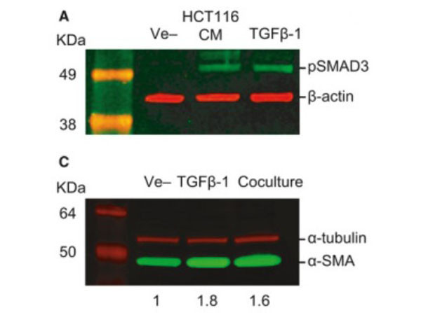
Western blot using Donkey Anti-Mouse IgG DyLight?680. TGF‐β‐mediated crosstalk between pericytes and CRC cells modulates pericyte secretome. (A) Incubation in HCT116 CM for 1?h induces SMAD3 phosphorylation in PC, as assessed by western blot. Exogenous recombinant TGF‐β (10?ng・mL−1) was used as a positive control, and β‐actin was used as loading control (n?=?3). (C) Western blot showing increased expression of αSMA in PC cocultured with HCT116 cells or stimulated with 10 ng・mL−1 TGF‐β1 for 48 h (n = 3). α‐tubulin was used as loading control. Numbers indicate the expression fold change relative to the loading control. Fig. 5. PMID: 32767843.
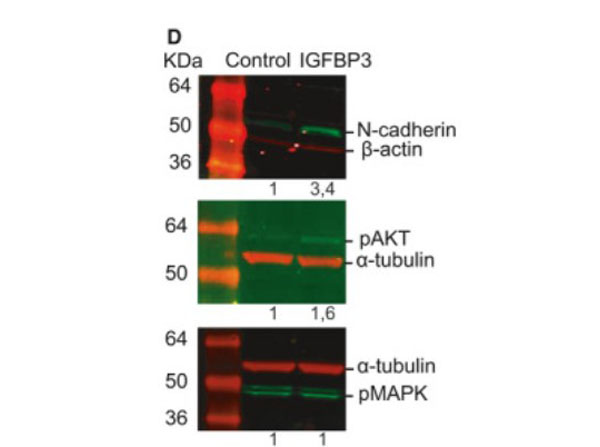
Western blot using Donkey Anti-Mouse IgG DyLight?680.Insulin‐like growth factor‐binding protein 3 increases CRC cell migration and invasion through Akt activation. (D) Treatment with 50?ng・mL−1?IGFBP‐3 for 72?h promotes the expression of N‐cadherin in HCT116 cells as assessed by western blot (top panel). Phosphorylation status of Akt (middle panel) and MAPK (bottom panel) in HCT116 cells treated with 50?ng・mL−1?IGFBP‐3 for 15?min. Representative images of three independent experiments (n?=?3). Numbers indicate the expression fold change relative to the loading control. Fig. 7. PMID: 32767843.
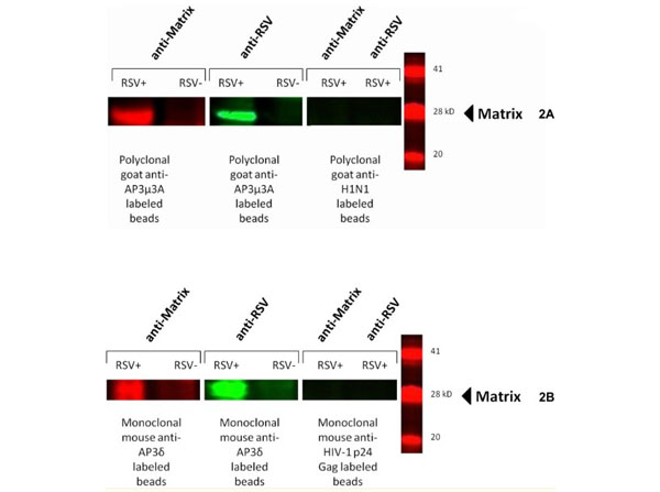
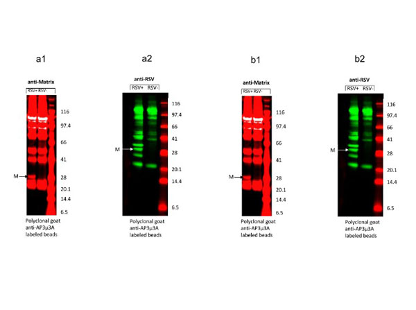
Western blot using Donkey Anti-Mouse IgG DyLight?680.The HRSV M protein co-immunoprecipitates with the AP-3Mu3A and AP-3delta complex during HRSV infection. HEp2 cells at approximately 90% confluency were either infected at an MOI of 5 or mock infected for 24 hours, cells were scraped or proteins were subsequently extracted using MPER. Cell lysates were incubated for 6 hours with 1 μg of either polyclonal goat anti-AP-3Mu3A or monoclonal mouse anti-AP-3delta along with a antibody specific isotype control, polyclonal goat-anti H1N1or monoclonal mouse anti-HIV-1 p24 Gag at 4°C on a rotating device. 20μl Protein A/G agarose beads were added to lysate plus corresponding antibody and incubated overnight. Immunoprecipate complex was pelleted and washed with PBS and then ran out on a SDS-PAGE gel and transferred to nitrocellulose membrane. Membrane was blocked and then probed with either monoclonal mouse anti-Matrix or polyclonal goat-anti HRSV primary antibody as described previously for one hour. Membranes were then washed with a PBS-Tween20 solution extensively and then probed with species-specific secondary antibodies donkey anti-goat DyLight800 and donkey anti-mouse DyLight680. Membranes were again washed extensively and blots were imaged on Odyssey Infrared imager. The results were reproducible in at least two independent assays. Fig 2. PMID: 29028839.
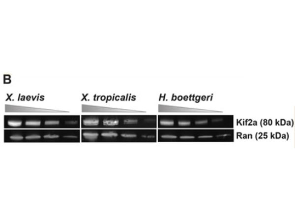
Western blot using Donkey Anti-Mouse IgG DyLight?680. Kif2a is enriched on spindles in?H. boettgeri?egg extracts, and inhibition of kif2a increases spindle length. B. Top Right Panel: Western blot of?X. laevis, X. tropicalis, and?H. boettgeri?extracts, probed for kif2a. Bottom Panel: Quantification of 3 separate blots for each species. Band intensities were normalized to the integrated density of the corresponding Ran loading control. AU=arbitrary units. Figure 3. PMID: 31630945.
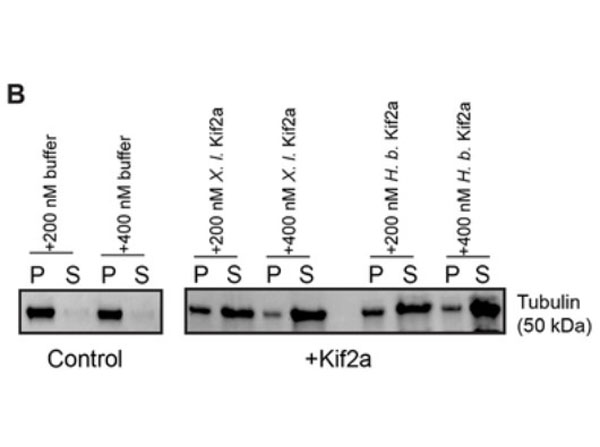
Serine 252 of kif2a regulates its activity.B. Top Panel: Increasing amounts of recombinant?X. laevis?or?H. boettgeri?kif2a proteins were added to taxol-stabilized microtubules and microtubules sedimented through a sucrose cushion. Amounts of soluble tubulin in the supernatant (S) and pellet (P) were quantified by SDS-PAGE and Coomassie staining. Bottom Panel: Ratio of pellet to supernatant gel band intensities in the microtubule sedimentation assay with 200 nM?H. boettgeri?or?X. laevis?kif2a added. Bands from 3 separate gels quantified, p=0.2768. NS= Not Significant. Error bars= +/− std dev. Figure 4. PMID: 31630945.
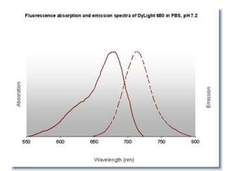
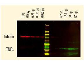
Properties of DyLight? Conjugates.
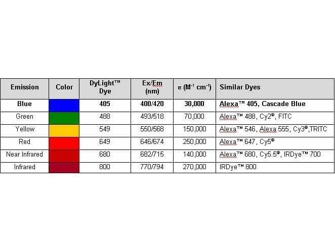
DyLight? dyes can be used for two-color western blot detection with low background and high signal. Anti-tubulin was detected using a DyLight? 680 conjugate. Anti-TNFa was detected using a DyLight? 800 conjugate. The image was captured using the OdysseyR Infrared Imaging System developed by LI-COR.
|
|
|
|
Western blot using Donkey Anti-Mouse IgG DyLight?680. TGF‐β‐mediated crosstalk between pericytes and CRC cells modulates pericyte secretome. (A) Incubation in HCT116 CM for 1?h induces SMAD3 phosphorylation in PC, as assessed by western blot. Exogenous recombinant TGF‐β (10?ng・mL−1) was used as a positive control, and β‐actin was used as loading control (n?=?3). (C) Western blot showing increased expression of αSMA in PC cocultured with HCT116 cells or stimulated with 10 ng・mL−1 TGF‐β1 for 48 h (n = 3). α‐tubulin was used as loading control. Numbers indicate the expression fold change relative to the loading control. Fig. 5. PMID: 32767843.
|
|
| 別品名 |
Donkey anti-Mouse IgG DyLight 680TM Conjugated Antibody, Donkey anti Mouse IgG Antibody DyLight 680TM Conjugation
|
| 交差種 |
Mouse
|
| 免疫動物 |
Donkey
|
| 標識物 |
DyLightTM 680
|
| 精製度 |
Affinity Purified
|
| 参考文献 |
[Pub Med ID]29028839
|
| [注意事項] |
濃度はロットによって異なる可能性があります。メーカーDS及びCoAからご確認ください。
|
|
| メーカー |
品番 |
包装 |
|
RKL
|
610-744-002
|
100 UG
|
※表示価格について
| 当社在庫 |
なし
|
| 納期目安 |
約10日
|
| 法規制 |
毒
|
| 保存温度 |
4℃
|
|
※当社では商品情報の適切な管理に努めておりますが、表示される法規制情報は最新でない可能性があります。
また法規制情報の表示が無いものは、必ずしも法規制に非該当であることを示すものではありません。
商品のお届け前に最新の製品法規制情報をお求めの際はこちらへお問い合わせください。
|
※当社取り扱いの試薬・機器製品および受託サービス・創薬支援サービス(納品物、解析データ等)は、研究用としてのみ販売しております。
人や動物の医療用・臨床診断用・食品用としては、使用しないように、十分ご注意ください。
法規制欄に体外診断用医薬品と記載のものは除きます。
|
|
※リンク先での文献等のダウンロードに際しましては、掲載元の規約遵守をお願いします。
|
|
※CAS Registry Numbers have not been verified by CAS and may be inaccurate.
|