|
※サムネイル画像をクリックすると拡大画像が表示されます。
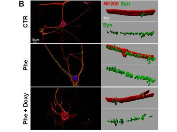
Synaptophysin density alterations in phenylalanine-treated hippocampal neurons. Hippocampal “sandwich” co-cultures were treated with Phe/Doxy for 72?h. Cells were double-stained with NF200 (red) and synaptophysin (green); nuclei were marked by Hoechst 33258 (blue). The number of synaptophysin-positive spots was normalized to the total volume of neurofilaments (NF200 expression) after three-dimensional reconstruction of the marker signals (representative images in (B). At least ten fields (1200x) for each condition from three independent experiments were analyzed. (A) The density of synaptophysin-positive spots was significantly increased by 20?mM Phe (*p?<?0.05 versus control; One-way ANOVA and Dunnett’s post-test). Treatment with 25?μM Doxy showed a trend towards a reduction in synaptophysin density, increasing towards control levels. Figure 11. PMID: 26510963.
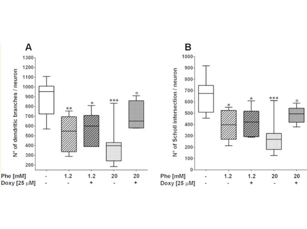
Phenylalanine-induced dendritic sprouting alterations in hippocampal neurons prevented by doxycycline co-treatment. Hippocampal “sandwich” co-cultures were treated with Phe/Doxy for 72?h. The neuron fractions were stained with NF200 and nuclei were marked by Hoechst 33258. At least eight fields (600x) for each condition from three independent experiments were analyzed. The number of dendritic branches (A) and Sholl intersections (B) was significantly decreased by Phe treatments. Treatment with 25?μM Doxy significantly counteracted the dendritic alterations induced by 20?mM Phe. *p?<?0.05, **p?<?0.01, ***p?<?0.001 versus control. °p?<?0.05?vs 20?mM Phe. One-way ANOVA and Tukey’s post-test. Figure 10. PMID: 26510963.
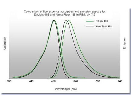
Properties of DyLight? Fluorescent Dyes.
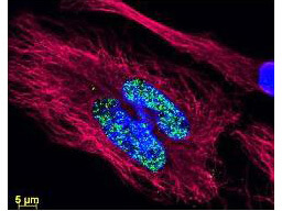
DyLight? dyes can be used for multi-color immunofluorescence microscopy with uniform fluorescence intensity throughout the image. DyLight? dyes are exceptionally bright and photostable and are optimized for microscopy and microarray detection methods. This image shows anti-histone detection using a DyLight? 488 conjugate (green). Anti-Tubulin was detected using a DyLight? 549 conjugate (red). Nuclei were counter-stained using DAPI (blue). The image was captured using an Axio Imager.Z1 (Zeiss Micro Imaging Inc).
|
|
|
|
Synaptophysin density alterations in phenylalanine-treated hippocampal neurons. Hippocampal “sandwich” co-cultures were treated with Phe/Doxy for 72?h. Cells were double-stained with NF200 (red) and synaptophysin (green); nuclei were marked by Hoechst 33258 (blue). The number of synaptophysin-positive spots was normalized to the total volume of neurofilaments (NF200 expression) after three-dimensional reconstruction of the marker signals (representative images in (B). At least ten fields (1200x) for each condition from three independent experiments were analyzed. (A) The density of synaptophysin-positive spots was significantly increased by 20?mM Phe (*p?<?0.05 versus control; One-way ANOVA and Dunnett’s post-test). Treatment with 25?μM Doxy showed a trend towards a reduction in synaptophysin density, increasing towards control levels. Figure 11. PMID: 26510963.
|
|
| 別品名 |
Donkey anti-Mouse IgG DyLight 488TM Conjugated Antibody, Donkey anti Mouse IgG Antibody DyLight 488TM Conjugation
|
| 交差種 |
Mouse
|
| 免疫動物 |
Donkey
|
| 標識物 |
DyLightTM 488
|
| 精製度 |
Affinity Purified
|
| 参考文献 |
[Pub Med ID]26510963
|
| [注意事項] |
濃度はロットによって異なる可能性があります。メーカーDS及びCoAからご確認ください。
|
|
| メーカー |
品番 |
包装 |
|
RKL
|
610-741-002
|
100 UG
|
※表示価格について
| 当社在庫 |
なし
|
| 納期目安 |
約10日
|
| 法規制 |
毒
|
| 保存温度 |
4℃
|
|
※当社では商品情報の適切な管理に努めておりますが、表示される法規制情報は最新でない可能性があります。
また法規制情報の表示が無いものは、必ずしも法規制に非該当であることを示すものではありません。
商品のお届け前に最新の製品法規制情報をお求めの際はこちらへお問い合わせください。
|
※当社取り扱いの試薬・機器製品および受託サービス・創薬支援サービス(納品物、解析データ等)は、研究用としてのみ販売しております。
人や動物の医療用・臨床診断用・食品用としては、使用しないように、十分ご注意ください。
法規制欄に体外診断用医薬品と記載のものは除きます。
|
|
※リンク先での文献等のダウンロードに際しましては、掲載元の規約遵守をお願いします。
|
|
※CAS Registry Numbers have not been verified by CAS and may be inaccurate.
|