
| 別品名 |
Cellular retinoic acid binding protein 2, RBP6, CRABP-II, Cellular retinoic acid binding protein 2, CRABP2, RBP6, CRABP-II, Cellular retinoic acid binding protein 2 Cellular retinoic acid binding protein II, CRABP II, CRABPII,
|
| 抗原部位 |
a.a.1-138
|
| 種由来 |
Human
|
| 標識物 |
Unlabeled
|
| 精製度 |
Ig fraction - Protein G
|
| 適用 |
Western Blot
Enzyme Linked Immunosorbent Assay
Immuno Fluorescence
Immunocytochemistry (cell)
|
| 免疫動物 |
Mouse
|
| 抗体クラス |
IgG2aκ
|
| クローン |
AT2E11
|
| 交差種 |
Human
|
| Accession No.(Gene/Protein) |
NP_001869, P29373
|
| 形状 |
液状
|
| 参考文献 |
Astrom,A., et al (1991). J. Biol. Chem. 266 (26), 17662-17666
Gupta,A., et al (2006). Cancer Res. 66 (16), 8100-8108
|
|
※サムネイル画像をクリックすると拡大画像が表示されます。
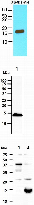
Cell lysates of mouse eye (60ug) were resolved by SDS-PAGE, transferred to NC membrane and probed with anti-human CRABP2 (1:250). Proteins were visualized using a goat anti-mouse secondary antibody conjugated to HRP and an ECL detection system.
The Cell lysates (40ug) were resolved by SDS-PAGE, transferred to PVDF membrane and probed with anti-human
CRABP2 antibody (1:250). Proteins were visualized using a goat anti-mouse secondary antibody conjugated to HRP and an ECL detection system.
Lane 1. : MCF-7 cell lysate
The Cell lysates (10ug) were resolved by SDS-PAGE, transferred to PVDF membrane and probed with anti-human CRABP2 antibody (1:1000). Proteins were visualized using a goat anti-mouse secondary antibody conjugated to HRP and an ECL detection system.
Lane 1. : 293T cell lysate
Lane 2. : CRABP2 Transfected 293T cell lysate
ICC/IF analysis of CRABP2 in HeLa cells line, stained with DAPI (Blue) for nucleus staining and monoclonal anti-human CRABP2 antibody (1:100) with goat anti-mouse IgG-Alexa fluor 488 conjugate (Green).
ICC/IF analysis of CRABP2 in MCF7 cells line, stained with DAPI (Blue) for nucleus staining and monoclonal anti-human CRABP2 antibody (1:100) with goat anti-mouse IgG-Alexa fluor 488 conjugate (Green).

|
|
|
|
Cell lysates of mouse eye (60ug) were resolved by SDS-PAGE, transferred to NC membrane and probed with anti-human CRABP2 (1:250). Proteins were visualized using a goat anti-mouse secondary antibody conjugated to HRP and an ECL detection system.
The Cell lysates (40ug) were resolved by SDS-PAGE, transferred to PVDF membrane and probed with anti-human
CRABP2 antibody (1:250). Proteins were visualized using a goat anti-mouse secondary antibody conjugated to HRP and an ECL detection system.
Lane 1. : MCF-7 cell lysate
The Cell lysates (10ug) were resolved by SDS-PAGE, transferred to PVDF membrane and probed with anti-human CRABP2 antibody (1:1000). Proteins were visualized using a goat anti-mouse secondary antibody conjugated to HRP and an ECL detection system.
Lane 1. : 293T cell lysate
Lane 2. : CRABP2 Transfected 293T cell lysate
|
|
|
| メーカー |
品番 |
包装 |
|
ATG
|
ATGA0134
|
50 UL
[1mg/ml]
|
※表示価格について
| 当社在庫 |
なし
|
| 納期目安 |
1週間程度
|
| 保存温度 |
-70℃
|
|

