|
※サムネイル画像をクリックすると拡大画像が表示されます。
Epifluorescence results of Donkey Anti-Rabbit IgG DyLight?405. Principle of Single Molecule Localization Microscopy (SMLM). A. Epifluorescence image of a single emitter, showing the ?200 nm width of the Point Spread Function (PSF, left panel) that is fitted using Gaussian curves (center panel) to determine its position with a ?20 nm precision (right panel). B. Epifluorescence image of microtubules in a COS cell. C. During SMLM acquisition, fluorescence emission is switched to a blinking mode and thousands of frames are recorded, showing individual blinking events that can be fitted to localize each emitter. D. After processing, all localizations are plotted to generate the SMLM images (bottom panels). Top panel is a zoom corresponding to the box highlighted in the full image and shows the gain in resolution with much thinner microtubules (top panels). Fig. 1. PMID: 31078795.

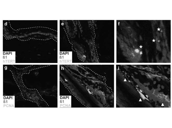
Immunofluorescence results using Donkey Anti-Rabbit IgG DyLight?405. After application of hTERT?DNA?PEI complex, hTERT protein expression promoted proliferation of hair follicle stem cells and enhanced hair growth. Immunofluorescent staining of hTERT and PCNA in dorsal skin treated with DNA?PEI complex (f, stars; i, triangles) at day 40 after wounding compared with non-treated control (d, g), respectively. (f, i) Displayed higher magnification micrographs of (e) and (h), respectively. Figure 6. PMID: 21593794.
Immunofluorescence results using Donkey Anti-Rabbit IgG DyLight?405.(e?f) The expression of hTERT (e) and PCNA (f) was detected by immunofluorescence microscopy using the Abs indicated. The transfected groups were shown as ‘D+P’ and non-transfected group were shown as ‘Neg’ at day 18 after transfection. The color reproduction of this figure is available on the html full text version of the manuscript. Figure 4. PMID: 21593794.

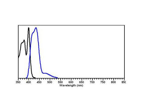
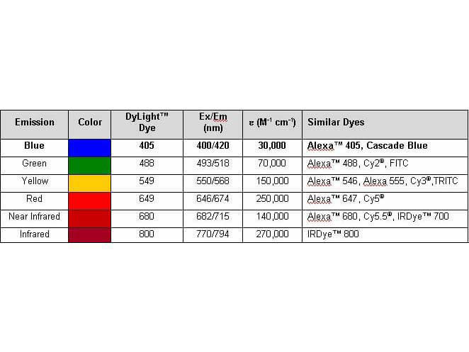
Properties of DyLight? Conjugates.
|
|
|
|
Epifluorescence results of Donkey Anti-Rabbit IgG DyLight?405. Principle of Single Molecule Localization Microscopy (SMLM). A. Epifluorescence image of a single emitter, showing the ?200 nm width of the Point Spread Function (PSF, left panel) that is fitted using Gaussian curves (center panel) to determine its position with a ?20 nm precision (right panel). B. Epifluorescence image of microtubules in a COS cell. C. During SMLM acquisition, fluorescence emission is switched to a blinking mode and thousands of frames are recorded, showing individual blinking events that can be fitted to localize each emitter. D. After processing, all localizations are plotted to generate the SMLM images (bottom panels). Top panel is a zoom corresponding to the box highlighted in the full image and shows the gain in resolution with much thinner microtubules (top panels). Fig. 1. PMID: 31078795.
|
|
| 別品名 |
Donkey Anti-Rabbit IgG Antibody DyLight 405TM Conjugated, Donkey Anti Rabbit IgG DyLight 405TM Conjugated Antibody
|
| 交差種 |
Rabbit
|
| 非交差(吸収処理)種 |
Human
Mouse
Rat
Bovine
Chicken
Sheep
Goat
Guinea Pig
Hamster
Equine
|
| 免疫動物 |
Donkey
|
| 標識物 |
DyLightTM 405
|
| 精製度 |
Affinity Purified
|
| 参考文献 |
[Pub Med ID]21593794
|
| [注意事項] |
濃度はロットによって異なる可能性があります。メーカーDS及びCoAからご確認ください。
|
|
| メーカー |
品番 |
包装 |
|
RKL
|
611-746-127
|
100 UG
|
※表示価格について
| 当社在庫 |
なし
|
| 納期目安 |
約10日
|
| 法規制 |
毒
|
| 保存温度 |
4℃
|
|
※当社では商品情報の適切な管理に努めておりますが、表示される法規制情報は最新でない可能性があります。
また法規制情報の表示が無いものは、必ずしも法規制に非該当であることを示すものではありません。
商品のお届け前に最新の製品法規制情報をお求めの際はこちらへお問い合わせください。
|
※当社取り扱いの試薬・機器製品および受託サービス・創薬支援サービス(納品物、解析データ等)は、研究用としてのみ販売しております。
人や動物の医療用・臨床診断用・食品用としては、使用しないように、十分ご注意ください。
法規制欄に体外診断用医薬品と記載のものは除きます。
|
|
※リンク先での文献等のダウンロードに際しましては、掲載元の規約遵守をお願いします。
|
|
※CAS Registry Numbers have not been verified by CAS and may be inaccurate.
|