|
※サムネイル画像をクリックすると拡大画像が表示されます。
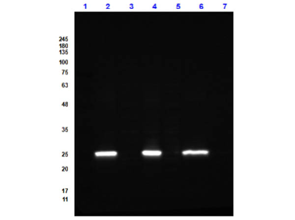
Western Bot of Goat Anti-GFP (GOAT) Antibody DyLight? 488 Conjugated. Lane 1: Opal Prestained Molecular Weight Marker (p/n MB-210-0500). Lane 2: GFP / HEK293T Whole Cell Lysate Reduced [0.05/10μg]. Lane 3: HEK293T Whole Cell Lysate Reduced (p/n W09-001-GX5) [10μg]. Lane 4: GFP / NIH-3T3 Whole Cell Lysate Reduced [0.05/10μg]. Lane 5: NIH-3T3 Whole Cell Lysate Reduced (p/n W10-000-358) [10μg]. Lane 6: GFP / PC-12 Whole Cell Lysate Reduced [0.05/10μg].Lane 7: PC-12 Whole Cell Lysate Reduced (p/n W12-001-GL9) [10μg]. Secondary Antibody: Goat Anti-GFP (GOAT) Antibody DyLight? 488 Conjugated at 1.0μg/mL overnight at 2-8°C. Blocking Buffer: Blocking Buffer for Fluorescent Western Blotting (p/n MB-070) for 1 hour at RT. Expect: 27kDa.
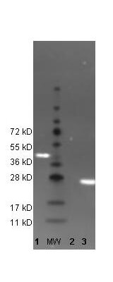
Western Blot of DyLight? 488 conjugated anti-GFP antibody to detect GFP control proteins. Lane 1: His-Sumo-GFP. Lane: Molecular Weight. Lane 2: Beta-Galactosidase (negative control). Lane 3: recombinant GFP control protein (000-001-215). Load: 35 μg per lane. Primary antibody: none. Secondary antibody: DyLight? 488 conjugated anti-GFP goat secondary antibody at 1:5,000. Block: MB-070 for 2 hr at RT. Predicted/Observed size: 27kDa/54kDa, 27kDa for rGFP/~45kDa His-Sumo-GFP. Other band(s): none.
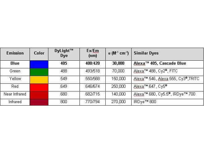
Properties of DyLight? Fluorescent Dyes. The emission spectra for this DyLight? conjugate match the principle output wavelengths of most common fluorescence instrumentation.
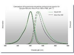
Comparison of fluorescence absorbtion and emission spectra for DyLight? 488 and Alexa Fluor 488 in PBS, pH7.2. The emission spectra for this DyLight? conjugate match the principle output wavelengths of most common fluorescence instrumentation.
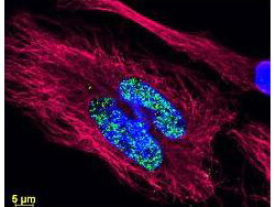
Multi-color Immunofluorescence Microscopy using DyLight? dyes. Tissue: human breast carcinoma. Fixation: 0.5% PFA. Antigen retrieval: not required. Primary antibody: Anti-Histone and Anti-Tubulin antibody at 10 μg/mL for 1 h at RT. Secondary antibody: DyLight? 488 conjugate and DyLight? 549 conjugate goat secondary antibody at 1:10,000 for 45 min at RT. Localization: Histone is nuclear and Tubulin is cytoplasmic. Staining: Anti-Histone detection using a DyLight? 488 conjugate (green) fluorescent signal and Anti-Tubulin was detected using a DyLight? 549 conjugate (red) fluorescent signal. Nuclei were counter-stained using DAPI (blue).
|
|
|
|
Western Bot of Goat Anti-GFP (GOAT) Antibody DyLight? 488 Conjugated. Lane 1: Opal Prestained Molecular Weight Marker (p/n MB-210-0500). Lane 2: GFP / HEK293T Whole Cell Lysate Reduced [0.05/10μg]. Lane 3: HEK293T Whole Cell Lysate Reduced (p/n W09-001-GX5) [10μg]. Lane 4: GFP / NIH-3T3 Whole Cell Lysate Reduced [0.05/10μg]. Lane 5: NIH-3T3 Whole Cell Lysate Reduced (p/n W10-000-358) [10μg]. Lane 6: GFP / PC-12 Whole Cell Lysate Reduced [0.05/10μg].Lane 7: PC-12 Whole Cell Lysate Reduced (p/n W12-001-GL9) [10μg]. Secondary Antibody: Goat Anti-GFP (GOAT) Antibody DyLight? 488 Conjugated at 1.0μg/mL overnight at 2-8°C. Blocking Buffer: Blocking Buffer for Fluorescent Western Blotting (p/n MB-070) for 1 hour at RT. Expect: 27kDa.
|
|
| 別品名 |
goat anti-GFP Antibody DyLightTM 488 Conjugation, DyLightTM 488 conjugated goat anti-GFP antibody, Green Fluorescent Protein, GFP antibody, Green Fluorescent Protein antibody, EGFP, enhanced Green Fluorescent Protein, Aequorea victoria, Jellyfish
|
| 非交差(吸収処理)種 |
Human
Mouse
Rat
|
| 適用 |
Western Blot
Dot Blot
|
| 免疫動物 |
Goat
|
| 標識物 |
DyLightTM 488
|
| 精製度 |
Affinity Purified
|
| Accession No.(Gene/Protein) |
P42212
|
| Tag情報 |
GFP
|
| 参考文献 |
[Pub Med ID]29288597
|
| [注意事項] |
濃度はロットによって異なる可能性があります。メーカーDS及びCoAからご確認ください。
|
|
| メーカー |
品番 |
包装 |
|
RKL
|
600-141-215
|
100 UG
|
※表示価格について
| 当社在庫 |
なし
|
| 納期目安 |
約10日
|
| 法規制 |
毒
|
| 保存温度 |
4℃
|
|
※当社では商品情報の適切な管理に努めておりますが、表示される法規制情報は最新でない可能性があります。
また法規制情報の表示が無いものは、必ずしも法規制に非該当であることを示すものではありません。
商品のお届け前に最新の製品法規制情報をお求めの際はこちらへお問い合わせください。
|
※当社取り扱いの試薬・機器製品および受託サービス・創薬支援サービス(納品物、解析データ等)は、研究用としてのみ販売しております。
人や動物の医療用・臨床診断用・食品用としては、使用しないように、十分ご注意ください。
法規制欄に体外診断用医薬品と記載のものは除きます。
|
|
※リンク先での文献等のダウンロードに際しましては、掲載元の規約遵守をお願いします。
|
|
※CAS Registry Numbers have not been verified by CAS and may be inaccurate.
|
