
| 別品名 |
RAC-PK-alpha, Protein kinase B, PKB, C-AKT, RAC-alpha serine/threonine-protein kinase, Proto-oncogene c-Akt, AKT1, AKT 1, AKT-1
|
| 種由来 |
Human
|
| 標識物 |
Unlabeled
|
| 精製度 |
Ig fraction - Protein A
|
| 適用 |
Western Blot
Immunohistochemistry
Immuno Fluorescence
|
| 免疫動物 |
Mouse
|
| 抗体クラス |
IgG1κ
|
| クローン |
17F6.B11
|
| 交差種 |
Human
Mouse
|
| 翻訳後修飾 |
リン酸化
|
| GENE ID |
207
|
| Accession No.(Gene/Protein) |
P31749
|
| Gene Symbol |
AKT1
|
| 形状 |
滅菌済み液状品
|
| 参考文献 |
[Pub Med ID]23913776
|
| [注意事項] |
濃度はロットによって異なる可能性があります。メーカーDS及びCoAからご確認ください。
|
|
※サムネイル画像をクリックすると拡大画像が表示されます。
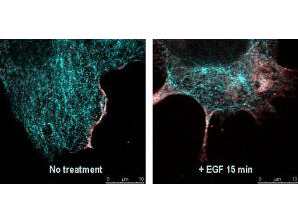
Immunofluorescence confocal microscopy of Mouse Anti AKT pS473 antibody. Tissue: EGF treated A431 cells. Fixation: 0.5% PFA. Antigen retrieval: EGF 15 min. Primary antibody: AKT pS473 antibody at 10 ug/mL for 1 h at RT. Secondary antibody: DyLight 488TM Goat anti Rabbit IgG, MAb anti AKT pS473, atto 647N anti Mouse IgG (Active Motif). at 1:10,000 for 45 min at RT. Localization: AKT pS473 is nuclear and occasionally cytoplasmic. Staining: AKT pS473 as red signal with tubulin (cyan).
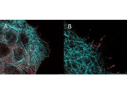
High resolution STED immunofluorescence nanoscopy of Mouse anti-AKT pS473 antibody. Tissue: A431 cells. The merge images (A) and at high magnification (B) show phosphorylated AKT colocalized with the distal microtubules. Fixation: 4% paraformaldehyde for 5 min and after washes blocked with 10% NGS/0.2% Triton X-100 for 30 min. Antigen retrieval: serum deprivation for 12 h. Primary antibody: AKT pS473 antibody at 10 ug/mL and α-tubulin (cyan) (p/n 600-401-880) at 1.4 ug/mL for 1 h at RT. Secondary antibody: Atto 647N anti-Mouse IgG (ATTO TEC GmbH), and DyLightTM488 anti-Rabbit IgG (p/n 611-141-122) were used at 1.0 ug/mL for 1h at RT for indirect detection. Localization: AKT pS473 is in the cytoplasm and also organized at the periphery of the cell. Staining: AKT pS473 as red signal with bis-benzimide (blue) nuclear counterstain.
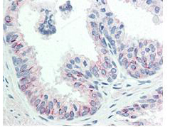
Immunohistochemistry of Mouse anti-AKT pS473 antibody. Tissue: human prostate tissue. Fixation: formalin fixed paraffin embedded. Antigen retrieval: not required. Primary antibody: AKT pS473 antibody at 20 ug/mL for 1 h at RT. Secondary antibody: Dako's Techmate streptavidin-biotin reagents at 1:10,000 for 45 min at RT. Localization: AKT pS473 is nuclear and occasionally cytoplasmic. Staining: AKT pS473 as precipitated red signal with hematoxylin purple nuclear counterstain.
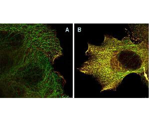
Immunofluorescence Microscopy of Mouse Anti-AKTpS473 antibody using STED nanoscopy to evaluate AKT activation and migration. Tissue: A431 cells. Antigen retrieval: Panel A: serum starved, unstimulated cells. Panel B: serum starved, EGF stimulated for 15 mins. A massive increase in AKT-pS473 activation, as measured by intensity signal, peaked at 15 minutes and was associated with depolymerized tubulin. Staining: Panel A shows STED data (AKT-pS473, red channel) collected simultaneously with confocal signal (a-tubulin, green channel). Upon stimulation of cells with EGF, a rapid activation of AKT is observed (Panel B) along with a coincident change in the tubulin organization (yellow signal), as well as an extensive cell shape-change (cell membrane folding) and accumulation of AKTpS473 at the cell periphery.
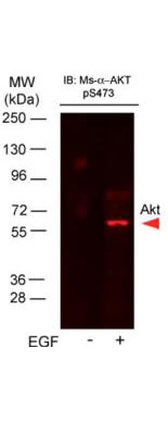
Western Blot of Mouse Anti-AKTpS473 antibody. Lane 1: A431 cell lysate (p/n W09-000-361). Lane 2: A431 cells stimulated for 15 min with EGF (p/n W09-000-362). Load: 35 ug per lane. Primary antibody: AKTpS473 antibody at 1:400 for overnight at 4C. Secondary antibody: DyLightTM649 Conjugated Anti-AKT pS473 Monoclonal Antibody (p/n 200-343-268) at 1:10,000 for 45 min at RT. Block: Blocking Buffer for Fluorescent Western Blotting (p/n MB-070) overnight at 4C. Predicted/Observed size: 56kDa. Other band(s): none.
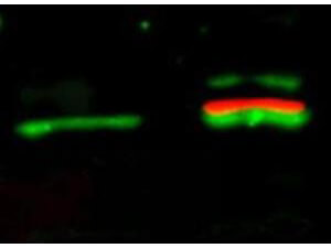
Western Blot of Mouse Anti-Akt pS473 antibody. Lane 1: unstimulated NIH/3T3 lysates (p/n W10-000-358) contain inactive unphosphorylated Akt1, green band. Lane 2: PDGF stimulated NIH/3T3 lysate (p/n W10-001-377) contains both inactive (green band) and activated phosphorylated Akt1 (red band). Load: 10 ug per lane. Primary antibody: rabbit anti-Akt (pan) (p/n 100-401-401) and mouse anti-Akt pS473 (p/n 200-301-B19) specific antibodies at 1:400 for overnight at 4C. Secondary antibody: anti-rabbit IgG DyLightTM 549 (green) and anti-mouse IgG DyLightTM 649 conjugated (red) secondary antibodies at 1:10,000 for 45 min at RT. Block: 5% BLOTTO overnight at 4C.
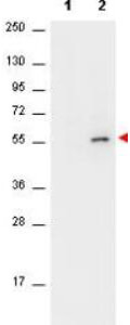
Western Blot of Mouse anti-AKT antibody. Lane 1: unstimulated NIH/3T3 cell lysates (p/n W10-000-358). Lane 2: PDGF stimulated NIH/3T3 cell lysates (p/n W10-001-377). Load: 10 ug per lane. Primary antibody: AKT antibody at 1:400 for overnight at 4C. Secondary antibody: HRP conjugated Gt-a-Mouse IgG (p/n 610-103-121) was used at a 1:40,000 dilution for 1 h at 4 C with FemtoMaxTM enhanced chemiluminescent reagent (p/n FEMTOMAX-100). Block: 5% BLOTTO (p/n B501-0500 in TBS for 2h at RT. Observed size: ~56 kDa for AKT. Other band(s): none.
|
|
|
|
Immunofluorescence confocal microscopy of Mouse Anti AKT pS473 antibody. Tissue: EGF treated A431 cells. Fixation: 0.5% PFA. Antigen retrieval: EGF 15 min. Primary antibody: AKT pS473 antibody at 10 ug/mL for 1 h at RT. Secondary antibody: DyLight 488TM Goat anti Rabbit IgG, MAb anti AKT pS473, atto 647N anti Mouse IgG (Active Motif). at 1:10,000 for 45 min at RT. Localization: AKT pS473 is nuclear and occasionally cytoplasmic. Staining: AKT pS473 as red signal with tubulin (cyan).
|
|
|
| メーカー |
品番 |
包装 |
|
RKL
|
200-301-B19
|
1 MG
|
※表示価格について
| 当社在庫 |
なし
|
| 納期目安 |
約10日
|
| 保存温度 |
-20℃
|
|

