|
※サムネイル画像をクリックすると拡大画像が表示されます。
Immunohistochemistry using Rockland Immunochemicals Anti-STAT5 pY694 monoclonal antibody shows detection of phosphorylated STAT5 pY694 in human breast tissue (40X). The antibody was used a dilution to 20 μg/mL. The image shows breast epithelium with moderate nuclear staining. Tissue was formalin fixed and paraffin embedded. No pre-treatment of sample was required. The image shows the localization of antibody as the precipitated red signal, with a hematoxylin purple nuclear counterstain. Personal communication, Andrew Elston, Lifespan Biosciences, Seattle, WA.

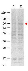
Western blot using Rockland's Protein A purified Mouse Monoclonal anti-Stat5 pY694 antibody shows detection of phosphorylated Stat5 (indicated by arrowhead at ~91 kDa) in NK92 cells after 30 min treatment with 1Ku of IL-2 (lane 2). No reactivity is seen for non-phosphorylated Stat5 in untreated cells (lane 1). The membrane was probed with the primary antibody at a 1:1,000 dilution, overnight at 4° C. For detection DyLight?800 conjugated Gt-a-Mouse IgG (p/n 610-145-002) was used at a 1:20,000 dilution for 30 min at room temperature followed by visualization using a VersaDoc? MP 4000 imaging system (Bio-Rad).
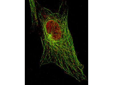
Immunofluorescent STED Microscopy of Anti-Stat5 pY694 (MOUSE) Monoclonal Antibody in 3T3 cells. Anti-Stat5 pY694 at 1:500 detecting Stat5 in 3T3 cells. Staining: Stat5 pY694 protein [Red]. Tubulin [Green].
|
|
|
|
Immunohistochemistry using Rockland Immunochemicals Anti-STAT5 pY694 monoclonal antibody shows detection of phosphorylated STAT5 pY694 in human breast tissue (40X). The antibody was used a dilution to 20 μg/mL. The image shows breast epithelium with moderate nuclear staining. Tissue was formalin fixed and paraffin embedded. No pre-treatment of sample was required. The image shows the localization of antibody as the precipitated red signal, with a hematoxylin purple nuclear counterstain. Personal communication, Andrew Elston, Lifespan Biosciences, Seattle, WA.
|
|
| 別品名 |
mouse anti-STAT5 pY694 Antibody, MGF antibody, Signal Transducer and Activator of Transcription 5A antibody, STAT 5 antibody
|
| 交差種 |
Human
Mouse
|
| 適用 |
Western Blot
Enzyme Linked Immunosorbent Assay
Immunohistochemistry
Immuno Fluorescence
|
| 免疫動物 |
Mouse
|
| クローン |
5F6.F1
|
| 抗体クラス |
IgG1κ
|
| 標識物 |
Unlabeled
|
| 精製度 |
Ig fraction - Protein A
|
| 翻訳後修飾 |
リン酸化
|
| GENE ID |
20850
|
| Accession No.(Gene/Protein) |
6755672, P42230
|
| Gene Symbol |
Stat5a
|
| 参考文献 |
Engblom,D., Kornfeld,J.W., Schwake,L., Tronche,F., Reimann,A., Beug,H., Hennighausen,L., Moriggl,R. and Schutz,G. (2007) Direct glucocorticoid receptor-Stat5 interaction in hepatocytes controls body size and maturation-related gene expression. Genes Dev. 21 (10), 1157-1162. Baugh,J.E. Jr., Floyd,Z.E. and Stephens,J.M. (2007) The modulation of STAT5A/GR complexes during fat cell differentiation and in mature adipocytes. Obesity (Silver Spring) 15 (3), 583-590. Laurence,A., Tato,C.M., Davidson,T.S., Kanno,Y., Chen,Z., Yao,Z., Blank,R.B., Meylan,F., Siegel,R., Hennighausen,L., Shevach,E.M. and O'Shea,J.J. (2007) Interleukin-2 signaling via STAT5 constrains T helper 17 cell generation. Immunity 26 (3), 371-381.
|
|
| メーカー |
品番 |
包装 |
|
RKL
|
200-301-B18
|
1 MG
|
※表示価格について
| 当社在庫 |
なし
|
| 納期目安 |
約10日
|
| 保存温度 |
-20℃
|
|
