|
※サムネイル画像をクリックすると拡大画像が表示されます。
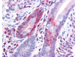
Rockland Antibody 200-301-964 has been tested in immunohistochemistry, analyzed by an anatomic pathologist and validated for use in IHC applications against formalin-fixed, paraffin-embedded human tissues. The antibody was serially diluted and tested at a range of concentrations on at least 22 different human formalin-fixed, paraffin archival tissues, and positive and negative tissues were scored and compared to the published literature on the expression and function of the gene. A representative image from positively stained small intestine shows the localization of the anti Pdcd4 antibody as the precipitated red signal, with a hematoxylin purple nuclear counterstain. Image provided courtesy of LifeSpan Biosciences, Seattle, WA
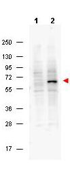
Western blot using Rockland's Protein A purified Mouse Monoclonal anti-Pdcd4 pS457 antibody shows detection of phosphorylated Pdcd4 (indicated by arrowhead at ~62 kDa) in NIH-3T3 cells (p/n W10-001-377) after 5 min treatment with 30 ng/mL PDGF [lane 2]. No reactivity is seen for (non-phosphorylated) in unstimulated NIH 3T3 cells (p/n W10-000-358) [lane 1]. The membrane was probed with the primary antibody at a 1:2,000 dilution, overnight at 4° C. For detection HRP conjugated Rb-a-Mouse IgG (p/n 610-4302) was used at a 1:20,000 dilution in blocking buffer (p/n MB-070) for 1 h at 4° C followed by visualization using a BiospectrumR imaging system (UVP).
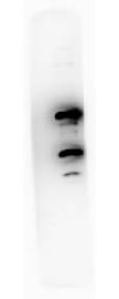
Western blot using Rockland Immunochemicals Protein A purified Mouse Monoclonal anti-Pdcd4 pS457 antibody against recombinant PDCD4 protein. Membrane was blocked in 1% BSA-TBS-T for 30 min RT and probed with 1° Ab Ms-A-Pdcd4pS457 1:1000 (o/n 4°C in 1% BSA-TBS-T) followed by 2° Ab Peroxidase Conjugated Rabbit anti-Ms CUST10M Lot# 20121 at 1:40,000 in MB-070 30 min RT. Bands at ~62 kD and ~32 kD were detected.
|
|
|
|
Rockland Antibody 200-301-964 has been tested in immunohistochemistry, analyzed by an anatomic pathologist and validated for use in IHC applications against formalin-fixed, paraffin-embedded human tissues. The antibody was serially diluted and tested at a range of concentrations on at least 22 different human formalin-fixed, paraffin archival tissues, and positive and negative tissues were scored and compared to the published literature on the expression and function of the gene. A representative image from positively stained small intestine shows the localization of the anti Pdcd4 antibody as the precipitated red signal, with a hematoxylin purple nuclear counterstain. Image provided courtesy of LifeSpan Biosciences, Seattle, WA
|
|
| 別品名 |
mouse anti-Pdcd4 pS457 Antibody, Death up-regulated gene protein antibody, Dug antibody, H731 antibody, Ma3 antibody, Neoplastic transformation inhibitor antibody, Neoplastic transformation inhibitor protein antibody, Nuclear antigen H731 antibody
|
| 交差種 |
Human
Mouse
|
| 適用 |
Western Blot
Enzyme Linked Immunosorbent Assay
Immunohistochemistry
|
| 免疫動物 |
Mouse
|
| クローン |
9G6
|
| 抗体クラス |
IgG1κ
|
| 標識物 |
Unlabeled
|
| 精製度 |
Ig fraction - Protein A
|
| 翻訳後修飾 |
リン酸化
|
| GENE ID |
27250
|
| Accession No.(Gene/Protein) |
21735596, Q53EL6
|
| Gene Symbol |
PDCD4
|
| 参考文献 |
Jansen AP, Camalier CE, Colburn NH (2005) Epidermal expression of the translation inhibitor programmed cell death 4 suppresses tumorigenesis. Cancer Res. 65(14):6034-6041. Palamarchuk A, Efanov A, Maximov V, Aqeilan RI, Croce CM, Pekarsky Y (2005) Akt phosphorylates and regulates Pdcd4 tumor suppressor protein. Cancer Res. 65 (24):11282-11286. Tzu-Hsuan Huang, GB Loeb, R Hsu, A Heidersbach, A Brincat, D Horiuchi, RJ Lebbink, YY Mo, A Goga, MT McManus. Up-regulation of miR-21 by HER2/neu Signaling Promotes Cell Invasion J. Biol. Chem. 2009 284: 18515-18524. First Published on May 6, 2009, doi:10.1074/jbc.M109.006676
|
| [注意事項] |
濃度はロットによって異なる可能性があります。メーカーDS及びCoAからご確認ください。
|
|
| メーカー |
品番 |
包装 |
|
RKL
|
200-301-964
|
100 UG
|
※表示価格について
| 当社在庫 |
なし
|
| 納期目安 |
約10日
|
| 保存温度 |
-20℃
|
|
※当社では商品情報の適切な管理に努めておりますが、表示される法規制情報は最新でない可能性があります。
また法規制情報の表示が無いものは、必ずしも法規制に非該当であることを示すものではありません。
商品のお届け前に最新の製品法規制情報をお求めの際はこちらへお問い合わせください。
|
※当社取り扱いの試薬・機器製品および受託サービス・創薬支援サービス(納品物、解析データ等)は、研究用としてのみ販売しております。
人や動物の医療用・臨床診断用・食品用としては、使用しないように、十分ご注意ください。
法規制欄に体外診断用医薬品と記載のものは除きます。
|
|
※リンク先での文献等のダウンロードに際しましては、掲載元の規約遵守をお願いします。
|
|
※CAS Registry Numbers have not been verified by CAS and may be inaccurate.
|
