|
※サムネイル画像をクリックすると拡大画像が表示されます。
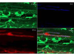
Immunohistochemistry of Rabbit Anti-Beta Actin Antibody. Tissue: sections 4.2 mm thick of rana pipiens tissue. Fixation: formalin fixed paraffin embedded. Antigen retrieval: not required. Primary antibody: Beta Actin antibody at 1:200 for 1 h at RT. Secondary antibody: Peroxidase rabbit secondary antibody at 1:10,000 for 45 min at RT. Localization: Beta Actin is at the neuromuscular junction. Staining: Beta Actin as precipitated green signal with red and blue counterstain.
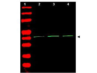
Western Blot of Rabbit Anti-Beta Actin Antibody. Lane 1: molecular weight. Lane 2: human embryonic kidney 293 (p/n W09-000-365). Lane 3: human lung carcinoma A549 (p/n W09-001-372). Lane 4: mouse brain (p/n W10-000-T004). Load: 35 μg per lane. Primary antibody: Beta Actin antibody at 1:1,500 for overnight at 4°C. Secondary antibody: IRDye800? rabbit secondary antibody at 1:10,000 for 45 min at RT. Block: 5% BLOTTO (p/n B501-0500) overnight at 4°C. Predicted/Observed size: ~42 kDa corresponding to beta Actin (arrowhead). Other band(s): none.
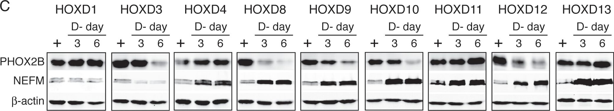
Distinct functions of HOXD proteins in regulation of neuronal differentiation.(A?B) Phase contrast imaging (A) and NEFM immunofluorescent staining (B) of BE(2)-C/Tet-Off/myc-HOXD cells cultured in the presence or absence of Doxy for 7 days. Scale bars, 50 μm. Shown are representative of three independent experiments with similar results. (C) Immunoblot analysis of PHOX2B and NEFM protein levels in the same cell samples described in Figure 3. Beta-actin levels are shown as loading control. Shown are representative of two independent experiments with similar results. (D) Quantification of NEFM mRNA levels in BE(2)-C/Tet-Off/myc-HOXD cells cultured in the presence or absence of Doxy for 3 days. The NEFM mRNA levels in BE(2)-C/Tet-Off/myc-HOXD cells in the presence of Doxy were designated as 1.0 (dashed line). The data were from two independent samples with each being assayed in triplicates and analyzed using two-tailed Student's t-test with the p values indicated. Error bars, SD. Figure provided by CiteAb. Source: PLoS One, PMID: 22879880.
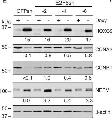
E2F6 is essential for HOXC9 induction of G1 arrest and repression of cell cycle genes. (A) Immunoblot analysis of E2F6 levels in BE(2)-C/Tet-Off/myc-HOXC9 cells infected with lentiviruses expressing shRNA against GFP or various coding regions of E2F6. E2F6 levels were quantified against β-actin. (B-D) Phase contrast imaging and growth assay (B) and cell cycle analysis (C, D) showing E2F6 knockdown abrogated HOXC9-induced growth arrest. Error bars, SD (n?=?4). (E-F) Immunoblot analysis (E) and quantification (F) showing that E2F6 knockdown abrogated HOXC9 repression of cyclins, but not HOXC9 induction of NEFM. HOXC9, CCNA2, CCNB1 and NEFM levels were quantified against β-actin with the protein levels in GFPsh-expressing cells cultured in the presence of doxycycline (Doxy+) were defined as 1.0 (dashed lines). Error bars, SD (n?=?3). Data in (D) and (F) were analyzed with unpaired, two-tailed Student’s t-test and p values are indicated. Figure provided by CiteAb. Source: BMC Genomics, PMID: 24274069.
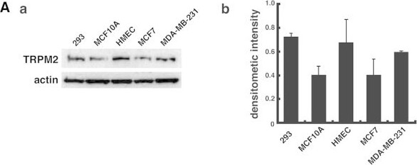
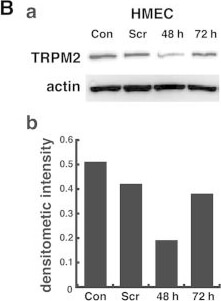
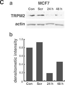
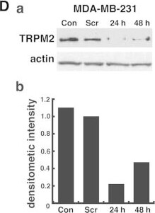
TRPM2 levels in breast cell lines and knockdown of TRPM2 levels by RNAi silencing. (A) Immunoblot (a) and densitometric quantification (b) of TRPM2 protein levels in two noncancerous breast cell lines (HMEC, MCF-10A) and two cancerous breast cell lines (MCF-7, MDA-MB-231). Immunoblot detection of TRPM2 protein levels in human embryonic kidney cells (HEK 293) provided the positive control. (B) RNAi silencing of TRPM2 in noncancerous HMEC cells (a), and quantification of protein levels by densitometry (b). (C and D) RNAi silencing of TRPM2 in MCF-7 and MDA-MB-231 breast adenocarcinoma cells (a) and quantification of protein levels by densitometry (b). All densitometric values represent TRPM2:actin ratios. Con, untreated negative control cells; Scr, treatment with scrambled negative control siRNA oligos. Figure provided by CiteAb. Source: Int J Oncol, PMID: 25760245.
TRPM2 levels in breast cell lines and knockdown of TRPM2 levels by RNAi silencing. (A) Immunoblot (a) and densitometric quantification (b) of TRPM2 protein levels in two noncancerous breast cell lines (HMEC, MCF-10A) and two cancerous breast cell lines (MCF-7, MDA-MB-231). Immunoblot detection of TRPM2 protein levels in human embryonic kidney cells (HEK 293) provided the positive control. (B) RNAi silencing of TRPM2 in noncancerous HMEC cells (a), and quantification of protein levels by densitometry (b). (C and D) RNAi silencing of TRPM2 in MCF-7 and MDA-MB-231 breast adenocarcinoma cells (a) and quantification of protein levels by densitometry (b). All densitometric values represent TRPM2:actin ratios. Con, untreated negative control cells; Scr, treatment with scrambled negative control siRNA oligos. Figure provided by CiteAb. Source: Int J Oncol, PMID: 25760245.
TRPM2 levels in breast cell lines and knockdown of TRPM2 levels by RNAi silencing. (A) Immunoblot (a) and densitometric quantification (b) of TRPM2 protein levels in two noncancerous breast cell lines (HMEC, MCF-10A) and two cancerous breast cell lines (MCF-7, MDA-MB-231). Immunoblot detection of TRPM2 protein levels in human embryonic kidney cells (HEK 293) provided the positive control. (B) RNAi silencing of TRPM2 in noncancerous HMEC cells (a), and quantification of protein levels by densitometry (b). (C and D) RNAi silencing of TRPM2 in MCF-7 and MDA-MB-231 breast adenocarcinoma cells (a) and quantification of protein levels by densitometry (b). All densitometric values represent TRPM2:actin ratios. Con, untreated negative control cells; Scr, treatment with scrambled negative control siRNA oligos. Figure provided by CiteAb. Source: Int J Oncol, PMID: 25760245.
TRPM2 levels in breast cell lines and knockdown of TRPM2 levels by RNAi silencing. (A) Immunoblot (a) and densitometric quantification (b) of TRPM2 protein levels in two noncancerous breast cell lines (HMEC, MCF-10A) and two cancerous breast cell lines (MCF-7, MDA-MB-231). Immunoblot detection of TRPM2 protein levels in human embryonic kidney cells (HEK 293) provided the positive control. (B) RNAi silencing of TRPM2 in noncancerous HMEC cells (a), and quantification of protein levels by densitometry (b). (C and D) RNAi silencing of TRPM2 in MCF-7 and MDA-MB-231 breast adenocarcinoma cells (a) and quantification of protein levels by densitometry (b). All densitometric values represent TRPM2:actin ratios. Con, untreated negative control cells; Scr, treatment with scrambled negative control siRNA oligos. Figure provided by CiteAb. Source: Int J Oncol, PMID: 25760245.
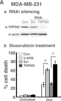
Effects of TRPM2 inhibition and RNAi knockdown in cancerous and non-cancerous breast cell lines after chemotherapeutic treatments. Immunoblot levels of TRPM2 protein after TRPM2 RNAi silencing (a) and quantification of cell death by flow cytometry (b) in (A) MDA-MB-231 breast adenocarcinoma, (B) MCF-7 breast adenocarcinoma and (C) human mammary epithelial cells (HMECs), a non-cancerous primary breast cell line. For RNAi silencing, cells were transfected with 100 nM anti-TRPM2 duplex siRNA oligos (TRPM2) or 100 nM negative control scrambled duplex siRNA oligos (Scr). Loading controls for immunoblots were provided by the immunodetection of β-actin. For chemotherapeutic treatments, the cells were pretreated 30 min with the TRPM2 inhibitor, 2-APB, and then subsequently treated with (A-b) 2 μM doxorubicin (Dox) until analysis; (B-b) 7 μM tamoxifen (Tam) until analysis; and (C-b) 2 μM doxorubicin (Dox) until analysis. Con, untransfected negative control cells. All error bars represent the SEM. *p<0.05 via one-way ANOVA and unpaired Student's t-test. Figure provided by CiteAb. Source: Oncol Rep, PMID: 26178079.
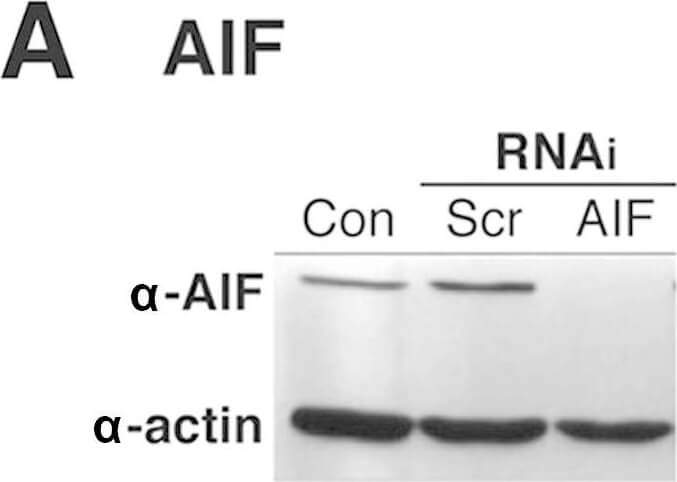
Analysis of poly(ADP-ribose)-mediated caspase-independent cell death in breast adenocarcinoma cells after TRPM2 inhibition and chemotherapeutic treatments. Immunoblot detection of (A) apoptosis-inducing factor (AIF) and (B) poly(ADP-ribose) glycohydrolase (PARG) in MDA-MB-231 breast adenocarcinoma cells after RNAi silencing. Loading controls for immunoblots were provided by the immunodetection of β-actin. Con, untransfected cells; Scr, cells transfected with negative control scrambled siRNA oligos. (C) Quantification of cell death by flow cytometry was performed in MDA-MB-231 cells after RNAi knockdown of AIF or PARG, pretreatment with 20 μM ACA for 30 min and treatment with 100 μM MNNG. *p<0.05, one-way ANOVA and unpaired Student's t-test; error bars represent the SEM. Figure provided by CiteAb. Source: Oncol Rep, PMID: 26178079.
|
|
|
|
Immunohistochemistry of Rabbit Anti-Beta Actin Antibody. Tissue: sections 4.2 mm thick of rana pipiens tissue. Fixation: formalin fixed paraffin embedded. Antigen retrieval: not required. Primary antibody: Beta Actin antibody at 1:200 for 1 h at RT. Secondary antibody: Peroxidase rabbit secondary antibody at 1:10,000 for 45 min at RT. Localization: Beta Actin is at the neuromuscular junction. Staining: Beta Actin as precipitated green signal with red and blue counterstain.
|
|
| 別品名 |
rabbit anti-beta Actin Antibody, Actin Antibody, Loading Control Antibody, beta actin, β actin, anti-beta actin antibody
|
| 交差種 |
Human
Mouse
|
交差種以外の
交差情報
(微交差など) |
[Reactivity]Leopard Frog
|
| 適用 |
Western Blot
Enzyme Linked Immunosorbent Assay
Immuno Fluorescence
|
| 免疫動物 |
Rabbit
|
| 抗原部位 |
a.a.359-368
|
| 標識物 |
Unlabeled
|
| 精製度 |
Affinity Purified
|
| Accession No.(Gene/Protein) |
NP_001092.1, P60709
|
| Gene Symbol |
ACTB
|
| 参考文献 |
[Pub Med ID]32132994
|
|
| メーカー |
品番 |
包装 |
|
RKL
|
600-401-886
|
200 UG
|
※表示価格について
| 当社在庫 |
なし
|
| 納期目安 |
約10日
|
| 保存温度 |
-20℃
|
|
