|
※サムネイル画像をクリックすると拡大画像が表示されます。
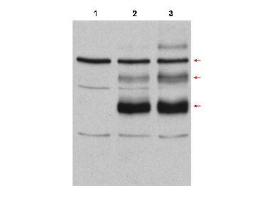
Western Blot of Rabbit anti-C-Myb antibody. Lane 1: Cos7 cell lysates vector. Lane 2: Cos7 cell lysates transfected with c-myb, no treatment. Lane 3: Cos7 cell lysates transfected with c-Myb and heat stress-treated (incubation at 42°C for 30 minutes) to induce c-Myb modification. Load: 35 μg per lane. Primary antibody: cMyb antibody at 1:500 for overnight at 4°C. Secondary antibody: IRDye800TM rabbit secondary antibody at 1:10,000 for 45 min at RT. Block: 10% BLOTTO overnight at 4°C. Predicted/Observed size: Lane 3 shows detection of bands at 75, 98, and 125kDa corresponding to overexpressed c-Myb (arrows): wild-type, with one attached SUMO molecule, and with two attached SUMO molecules, respectively.
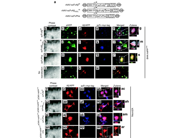
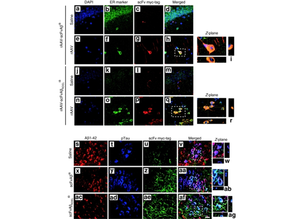
AAV vector?mediated expression of the Aβ-specific intrabodies in 3xTg-AD mice reveals the intrabodies alter general patterns of cell-associated Aβ42. Two month-old 3xTg-AD mice were unilaterally injected in the CA1 region of the hippocampus with saline (a?d?and?j?m), recombinant adeno-associated virus (rAAV)-scFvAβIB?(e?i) and rAAV-scFvAβKDELIB?(n?r). Nine months postinjection animals were killed and brain sections were processed for coimmunocytochemistry with the following: 4',6-diamidino-2-phenylindole (blue;?a,e,j,n), endoplasmic reticulum-specific antibody (green;?b,f,k,o) and c-myc?epitope tag-specific antibody (red;?c,g,l,p). Co-registered staining is indicated in the merged images as yellow (d,h,m,q). Panels?i?and?r?represent digitally magnified images, which illustrate a cross section through the?Z-plane obtained with confocal microscopy. All images were obtained at ×100 original magnification. Additional brain sections from these rAAV vector-injected 3xTg-AD mice were processed for fluorescence coimmunocytochemistry with the following: Aβ42-specific antibody (red;?s,x,ac), phospho-Tau (AT180)-specific antibody (blue;?t,y,ad) and c-myc?epitope tag-specific antibody (green;?u,z,ae). Co-registered staining for all markers is indicated in the merged images as white or for Aβ42 and phospho-Tau as purple (v,w,aa,ab,af,ag). Panels?w,ab, and?ag?represent digitally magnified images, which illustrate a cross section through the?Z-plane obtained with confocal microscopy. All images were obtained at ×100 original magnification. scFv, single-chain variable fragment; Aβ, amyloid-β; IB, intrabody. Fig 4. PMID:?19638957
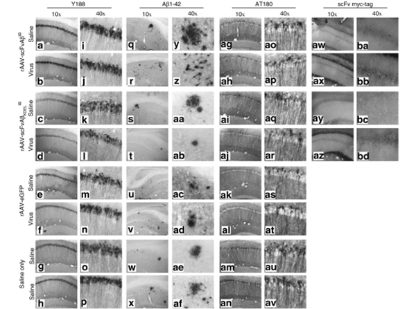
Qualitative effects of chronic anti-Aβ42 intrabody expression on AD-related pathological hallmarks.?Two month-old 3xTg-Alzheimer's disease mice were unilaterally injected in the CA1 region of the hippocampus with saline, recombinant adeno-associated virus (rAAV)-enhanced green fluorescent protein, rAAV-scFvAβIB?or rAAV-scFvAβKDELIB. Nine months postinjection animals were killed and 30-μm brain sections were processed for immunohistochemical analyses of hAPP transgene expression (Y188 antibody;?a?p), extracellular Aβ 1?42 deposition (anti-Aβ42 antibody;?q?af), hyperphosphorylated Tau (AT180 antibody;?ag?av), and c-myc?epitope tag-specific antibody (aw?bd). Of note, the images obtained for the c-myc?epitope tag immunohistochemical analysis were obtained originally in color by fluorescence microscopy, converted to black-and-white, and subsequently inverted to provide comparable views to the other sections captured using conventional light microscopy following 3,3'-diaminobenzidine immunohistochemistry. Representative images of the infused hippocampus are displayed at ×10 (a?h,q?x,ag?an,aw?az) and ×40 original magnification (i?p,y?af,ao?av,ba?bd). scFv, single-chain variable fragment; Aβ, amyloid-β; IB, intrabody. Fig 5. PMID:?19638957
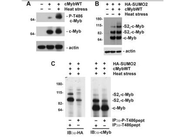
Phosphorylation of Thr486?negatively regulates stress-induced SUMOylation of c-Myb.?A). COS7 cells were transfected with a construct encoding c-MybWT and treated with hyperthermia as indicated. Immunoblotting results are of total cell lysates analyzed with rabbit polyclonal antibody specific for phosphorylated Thr486?(P-cMybT486 p/n 600-401-C47,?top panel), nonphosphorylated Thr486?in murine c-Myb (c-Myb p/n 600-401-B06,?middle panel), or anti-actin antibody (bottom panel).?B). cell lysates from COS7 cells transfected with construct encoding c-MybWT and HA-SUMO-2 and treated with hyperthermia were analyzed by immunoblotting with monoclonal anti-c-Myb (top panel), or anti-actin antibody (bottom panel). The mobility of unmodified (c-Myb) and SUMOylated forms (HAS21-cMyb and HAS22-cMyb) are marked.?C). immunoprecipitation of c-Myb using rabbit polyclonal antibody specific for phosphorylated (α-P-T486pept p/n 600-401-C47) or nonphosphorylated Thr486?(α-T486pept p/n 600-401-B06) were analyzed by immunoblotting with monoclonal anti-HA (α-HA,?left panel) or anti-c-Myb antibody (α-cMyb,?right panel).?Fig 3. PMID:?24257756
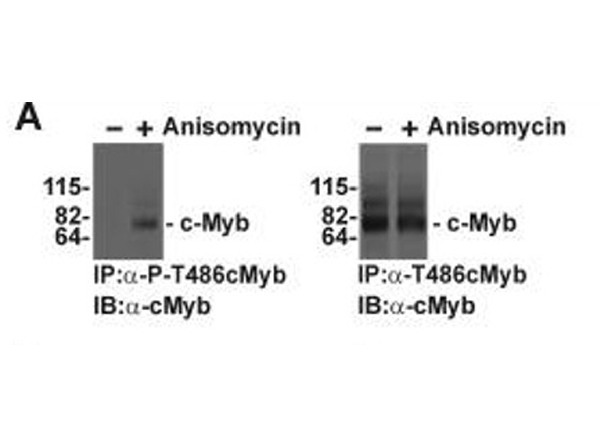
Regulation of endogenous c-Myb SUMOylation by p38 MAPKs in myeloid cells.?A, M1 cells expressing endogenous c-Myb and SUMO-2/3 were treated with anisomycin for 20 min as indicated, and c-Myb was immunoprecipitated with rabbit polyclonal antibody specific for phosphorylated (IP:α-P-T486cMyb, p/n 600-401-C47 left panel) or nonphosphorylated Thr486?(IP:α-T486cMyb,?p/n 600-401-B06 right panel). Immunoprecipitates were analyzed by immunoblotting with monoclonal anti-c-Myb antibody (IB:α-cMyb). Fig 5. PMID:?24257756
|
|
|
|
Western Blot of Rabbit anti-C-Myb antibody. Lane 1: Cos7 cell lysates vector. Lane 2: Cos7 cell lysates transfected with c-myb, no treatment. Lane 3: Cos7 cell lysates transfected with c-Myb and heat stress-treated (incubation at 42°C for 30 minutes) to induce c-Myb modification. Load: 35 μg per lane. Primary antibody: cMyb antibody at 1:500 for overnight at 4°C. Secondary antibody: IRDye800TM rabbit secondary antibody at 1:10,000 for 45 min at RT. Block: 10% BLOTTO overnight at 4°C. Predicted/Observed size: Lane 3 shows detection of bands at 75, 98, and 125kDa corresponding to overexpressed c-Myb (arrows): wild-type, with one attached SUMO molecule, and with two attached SUMO molecules, respectively.
|
|
| 別品名 |
rabbit anti-c-Myb Antibody, Proto-oncogene c-Myb antibody, C myb antibody, MYB antibody, Transcriptional activator Myb, cmyb antibody
|
| 別包装 |
あり
|
| 交差種 |
Mouse
Monkey
|
| 適用 |
Western Blot
Enzyme Linked Immunosorbent Assay
|
| 免疫動物 |
Rabbit
|
| 標識物 |
Unlabeled
|
| 精製度 |
Affinity Purified
|
| GENE ID |
17863
|
| Accession No.(Gene/Protein) |
110556654, P06876
|
| Gene Symbol |
Myb
|
| 参考文献 |
[[Cosmo Reference Collection]][Pub Med ID]19638957
|
| [注意事項] |
濃度はロットによって異なる可能性があります。メーカーDS及びCoAからご確認ください。
|
|
| メーカー |
品番 |
包装 |
|
RKL
|
600-401-B06
|
100 UG
|
※表示価格について
| 当社在庫 |
なし
|
| 納期目安 |
約10日
|
| 保存温度 |
-20℃
|
|


