|
※サムネイル画像をクリックすると拡大画像が表示されます。
Figure 1. Western blot analysis of YWHAE using anti-YWHAE antibody (M01687-2).
Electrophoresis was performed on a 5-20% SDS-PAGE gel at 70V (Stacking gel) / 90V (Resolving gel) for 2-3 hours. The sample well of each lane was loaded with 30 ug of sample under reducing conditions.
Lane 1: human Hela whole cell lysates,
Lane 2: human SH-SY5Y whole cell lysates,
Lane 3: human SW620 whole cell lysates,
Lane 4: human Raji whole cell lysates,
Lane 5: rat brain tissue lysates,
Lane 6: rat PC-12 whole cell lysates,
Lane 7: mouse brain tissue lysates,
Lane 8: mouse NIH/3T3 whole cell lysates.
After electrophoresis, proteins were transferred to a nitrocellulose membrane at 150 mA for 50-90 minutes. Blocked the membrane with 5% non-fat milk/TBS for 1.5 hour at RT. The membrane was incubated with mouse anti-YWHAE antigen affinity purified monoclonal antibody (Catalog # M01687-2) at 0.5 μg/mL overnight at 4°C, then washed with TBS-0.1%Tween 3 times with 5 minutes each and probed with a goat anti-mouse IgG-HRP secondary antibody at a dilution of 1:10000 for 1.5 hour at RT. The signal is developed using an Enhanced Chemiluminescent detection (ECL) kit (Catalog # EK1001) with Tanon 5200 system. A specific band was detected for YWHAE at approximately 29 kDa. The expected band size for YWHAE is at 29 kDa.
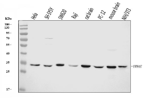
Figure 2. IHC analysis of YWHAE using anti-YWHAE antibody (M01687-2).
YWHAE was detected in a paraffin-embedded section of human colorectal adenocarcinoma tissue. Heat mediated antigen retrieval was performed in EDTA buffer (pH 8.0, epitope retrieval solution). The tissue section was blocked with 10% goat serum. The tissue section was then incubated with 2 μg/ml mouse anti-YWHAE Antibody (M01687-2) overnight at 4°C. Peroxidase Conjugated Goat Anti-mouse IgG was used as secondary antibody and incubated for 30 minutes at 37°C. The tissue section was developed using HRP Conjugated Mouse IgG Super Vision Assay Kit (Catalog # SV0001) with DAB as the chromogen.
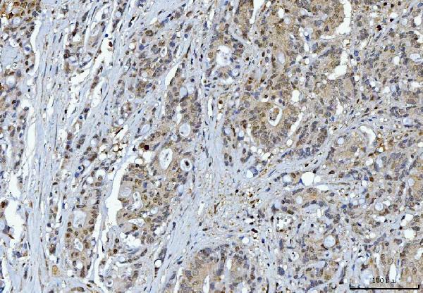
Figure 3. IHC analysis of YWHAE using anti-YWHAE antibody (M01687-2).
YWHAE was detected in a paraffin-embedded section of human liver cancer tissue. Heat mediated antigen retrieval was performed in EDTA buffer (pH 8.0, epitope retrieval solution). The tissue section was blocked with 10% goat serum. The tissue section was then incubated with 2 μg/ml mouse anti-YWHAE Antibody (M01687-2) overnight at 4°C. Peroxidase Conjugated Goat Anti-mouse IgG was used as secondary antibody and incubated for 30 minutes at 37°C. The tissue section was developed using HRP Conjugated Mouse IgG Super Vision Assay Kit (Catalog # SV0001) with DAB as the chromogen.
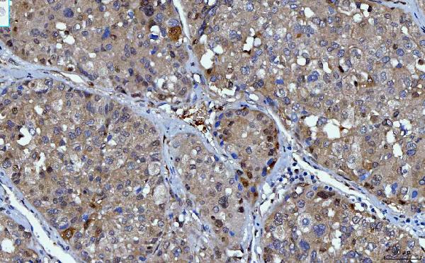
Figure 4. IHC analysis of YWHAE using anti-YWHAE antibody (M01687-2).
YWHAE was detected in a paraffin-embedded section of human tonsil tissue. Heat mediated antigen retrieval was performed in EDTA buffer (pH 8.0, epitope retrieval solution). The tissue section was blocked with 10% goat serum. The tissue section was then incubated with 2 μg/ml mouse anti-YWHAE Antibody (M01687-2) overnight at 4°C. Peroxidase Conjugated Goat Anti-mouse IgG was used as secondary antibody and incubated for 30 minutes at 37°C. The tissue section was developed using HRP Conjugated Mouse IgG Super Vision Assay Kit (Catalog # SV0001) with DAB as the chromogen.
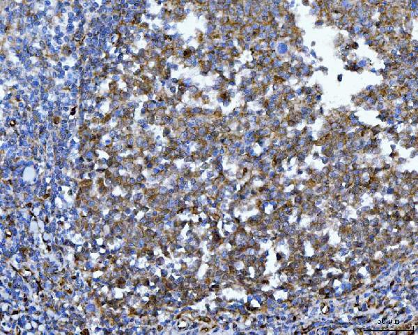
Figure 5. IHC analysis of YWHAE using anti-YWHAE antibody (M01687-2).
YWHAE was detected in a paraffin-embedded section of human endometrial cancer tissue. Heat mediated antigen retrieval was performed in EDTA buffer (pH 8.0, epitope retrieval solution). The tissue section was blocked with 10% goat serum. The tissue section was then incubated with 2 μg/ml mouse anti-YWHAE Antibody (M01687-2) overnight at 4°C. Peroxidase Conjugated Goat Anti-mouse IgG was used as secondary antibody and incubated for 30 minutes at 37°C. The tissue section was developed using HRP Conjugated Mouse IgG Super Vision Assay Kit (Catalog # SV0001) with DAB as the chromogen.
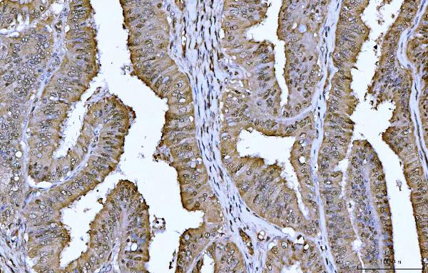
Figure 6. IHC analysis of YWHAE using anti-YWHAE antibody (M01687-2).
YWHAE was detected in a paraffin-embedded section of human placenta tissue. Heat mediated antigen retrieval was performed in EDTA buffer (pH 8.0, epitope retrieval solution). The tissue section was blocked with 10% goat serum. The tissue section was then incubated with 2 μg/ml mouse anti-YWHAE Antibody (M01687-2) overnight at 4°C. Peroxidase Conjugated Goat Anti-mouse IgG was used as secondary antibody and incubated for 30 minutes at 37°C. The tissue section was developed using HRP Conjugated Mouse IgG Super Vision Assay Kit (Catalog # SV0001) with DAB as the chromogen.
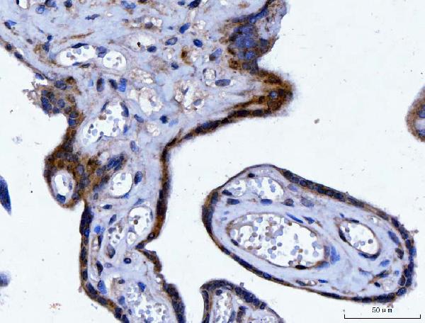
Figure 7. IHC analysis of YWHAE using anti-YWHAE antibody (M01687-2).
YWHAE was detected in a paraffin-embedded section of human bladder epithelial carcinoma tissue. Heat mediated antigen retrieval was performed in EDTA buffer (pH 8.0, epitope retrieval solution). The tissue section was blocked with 10% goat serum. The tissue section was then incubated with 2 μg/ml mouse anti-YWHAE Antibody (M01687-2) overnight at 4°C. Peroxidase Conjugated Goat Anti-mouse IgG was used as secondary antibody and incubated for 30 minutes at 37°C. The tissue section was developed using HRP Conjugated Mouse IgG Super Vision Assay Kit (Catalog # SV0001) with DAB as the chromogen.

Figure 8. IHC analysis of YWHAE using anti-YWHAE antibody (M01687-2).
YWHAE was detected in a paraffin-embedded section of human laryngeal squamous cell carcinomas tissue. Heat mediated antigen retrieval was performed in EDTA buffer (pH 8.0, epitope retrieval solution). The tissue section was blocked with 10% goat serum. The tissue section was then incubated with 2 μg/ml mouse anti-YWHAE Antibody (M01687-2) overnight at 4°C. Peroxidase Conjugated Goat Anti-mouse IgG was used as secondary antibody and incubated for 30 minutes at 37°C. The tissue section was developed using HRP Conjugated Mouse IgG Super Vision Assay Kit (Catalog # SV0001) with DAB as the chromogen.
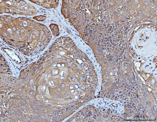
Figure 9. IF analysis of YWHAE using anti-YWHAE antibody (M01687-2).
YWHAE was detected in an immunocytochemical section of Caco-2 cells. Enzyme antigen retrieval was performed using IHC enzyme antigen retrieval reagent (AR0022) for 15 mins. The cells were blocked with 10% goat serum. And then incubated with 5 μg/mL mouse anti-YWHAE Antibody (M01687-2) overnight at 4°C. DyLightR594 Conjugated Goat Anti-Mouse IgG (BA1141) was used as secondary antibody at 1:100 dilution and incubated for 30 minutes at 37°C. The section was counterstained with DAPI. Visualize using a fluorescence microscope and filter sets appropriate for the label used.
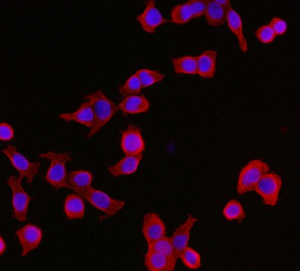
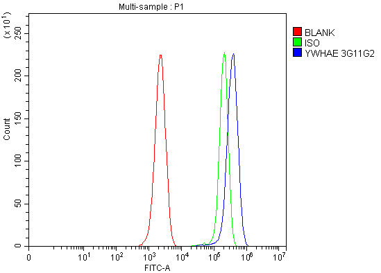
Figure 10. Flow Cytometry analysis of ANA-1 cells using anti-YWHAE antibody (M01687-2).
Overlay histogram showing ANA-1 cells stained with M01687-2 (Blue line). The cells were blocked with 10% normal goat serum. And then incubated with mouse anti-YWHAE Antibody (M01687-2, 1 μg/1x106 cells) for 30 min at 20°C. DyLightR488 conjugated goat anti-mouse IgG (BA1126, 5-10 μg/1x106 cells) was used as secondary antibody for 30 minutes at 20°C. Isotype control antibody (Green line) was mouse IgG (1 μg/1x106) used under the same conditions. Unlabelled sample (Red line) was also used as a control.
|
|
|
|
Figure 1. Western blot analysis of YWHAE using anti-YWHAE antibody (M01687-2).
Electrophoresis was performed on a 5-20% SDS-PAGE gel at 70V (Stacking gel) / 90V (Resolving gel) for 2-3 hours. The sample well of each lane was loaded with 30 ug of sample under reducing conditions.
Lane 1: human Hela whole cell lysates,
Lane 2: human SH-SY5Y whole cell lysates,
Lane 3: human SW620 whole cell lysates,
Lane 4: human Raji whole cell lysates,
Lane 5: rat brain tissue lysates,
Lane 6: rat PC-12 whole cell lysates,
Lane 7: mouse brain tissue lysates,
Lane 8: mouse NIH/3T3 whole cell lysates.
After electrophoresis, proteins were transferred to a nitrocellulose membrane at 150 mA for 50-90 minutes. Blocked the membrane with 5% non-fat milk/TBS for 1.5 hour at RT. The membrane was incubated with mouse anti-YWHAE antigen affinity purified monoclonal antibody (Catalog # M01687-2) at 0.5 μg/mL overnight at 4°C, then washed with TBS-0.1%Tween 3 times with 5 minutes each and probed with a goat anti-mouse IgG-HRP secondary antibody at a dilution of 1:10000 for 1.5 hour at RT. The signal is developed using an Enhanced Chemiluminescent detection (ECL) kit (Catalog # EK1001) with Tanon 5200 system. A specific band was detected for YWHAE at approximately 29 kDa. The expected band size for YWHAE is at 29 kDa.
|
|
| 別品名 |
14-3-3 protein epsilon, 14-3-3E, YWHAE
|
| 種由来 |
Human
|
| 交差種 |
Human
Mouse
Rat
|
| 適用 |
Western Blot
Immunohistochemistry
Immuno Fluorescence
Immunocytochemistry (cell)
Flow Cytometry
|
| 免疫動物 |
Mouse
|
| クローン |
3G11G2
|
| 抗原部位 |
a.a.1-255
|
| 精製度 |
Affinity Purified
|
| GENE ID |
7531
|
| Accession No.(Gene/Protein) |
P62258
|
| 分子量 |
29174
|
|
| メーカー |
品番 |
包装 |
|
ABH
|
ABO16243
|
100 UG
|
※表示価格について
| 当社在庫 |
なし
|
| 納期目安 |
2週間程度
|
| 保存温度 |
-20℃
|
|
※当社では商品情報の適切な管理に努めておりますが、表示される法規制情報は最新でない可能性があります。
また法規制情報の表示が無いものは、必ずしも法規制に非該当であることを示すものではありません。
商品のお届け前に最新の製品法規制情報をお求めの際はこちらへお問い合わせください。
|
※当社取り扱いの試薬・機器製品および受託サービス・創薬支援サービス(納品物、解析データ等)は、研究用としてのみ販売しております。
人や動物の医療用・臨床診断用・食品用としては、使用しないように、十分ご注意ください。
法規制欄に体外診断用医薬品と記載のものは除きます。
|
|
※リンク先での文献等のダウンロードに際しましては、掲載元の規約遵守をお願いします。
|
|
※CAS Registry Numbers have not been verified by CAS and may be inaccurate.
|
