|
※サムネイル画像をクリックすると拡大画像が表示されます。
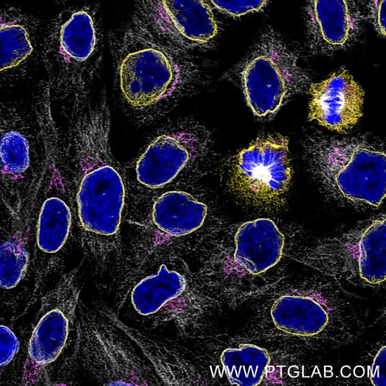
Immunofluorescence of Hela cells: PFA-fixed cells were stained with rabbit IgG anti-Lamin antibody (12987-1-AP) labeled with FlexAble 2.0 CoraLite Plus 555 Kit (KFA502, yellow), rabbit IgG anti-GM130 antibody (11308-1-AP) labeled with FlexAble 2.0 CoraLite Plus 647 Kit (KFA503, magenta) and rabbit IgG anti-Tubulin antibody (80713-1-RR) labeled with FlexAble 2.0 CoraLite Plus 750 Kit (KFA504, grey). Cell nuclei were stained with DAPI (blue). Confocal images were acquired with a 63x oil objective and post-processed. Images were recorded at the Core Facility Bioimaging at the Biomedical Center, LMU Munich.
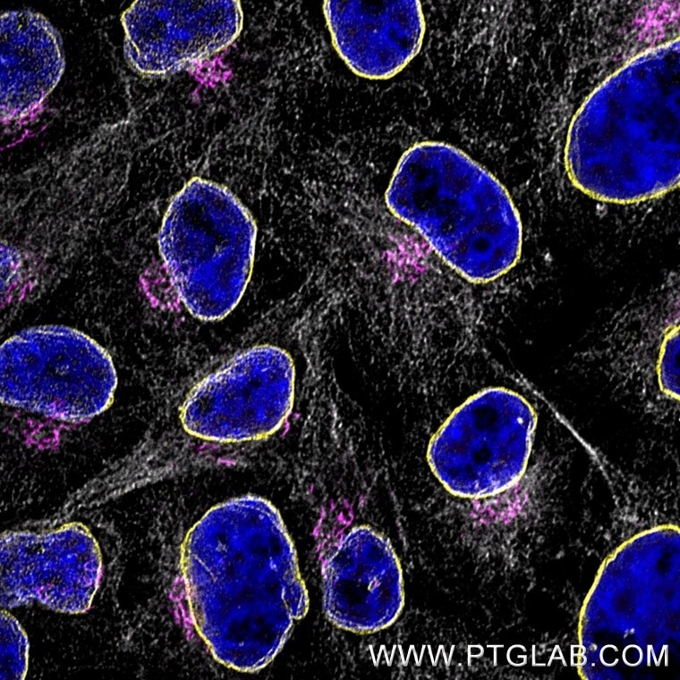
Immunofluorescence of Hela cells: PFA-fixed cells were stained with rabbit IgG anti-Lamin antibody (12987-1-AP) labeled with FlexAble 2.0 CoraLite Plus 555 Kit (KFA502, yellow), rabbit IgG anti-GM130 antibody (11308-1-AP) labeled with FlexAble 2.0 CoraLite Plus 647 Kit (KFA503, magenta) and rabbit IgG anti-Tubulin antibody (80713-1-RR) labeled with FlexAble 2.0 CoraLite Plus 750 Kit (KFA504, grey). Cell nuclei were stained with DAPI (blue). Confocal images were acquired with a 63x oil objective and post-processed. Images were recorded at the Core Facility Bioimaging at the Biomedical Center, LMU Munich.
Immunofluorescence of Hela cells: PFA-fixed cells were stained with rabbit IgG anti-TDP43 antibody directly conjugated with CoraLite Plus 488 (CL488-10782, green), rabbit IgG anti-GM130 antibody directly conjugated with CoraLite Plus 555 (CL555-11308, red), rabbit IgG anti-Lamin antibody (12987-1-AP) labeled with FlexAble 2.0 CoraLite Plus 647 Kit (KFA503, magenta) and rabbit IgG anti-Tubulin antibody (80713-1-RR) labeled with FlexAble 2.0 CoraLite Plus 750 Kit (KFA504, cyan).
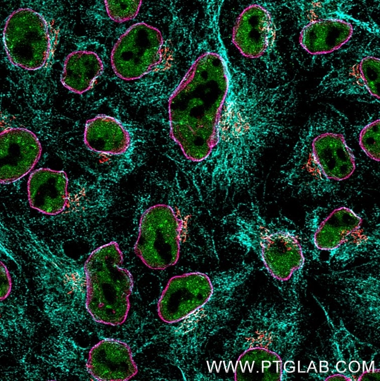
Immunofluorescence of Hela cells: PFA-fixed cells were stained with rabbit IgG anti-TDP43 antibody directly conjugated with CoraLite Plus 488 (CL488-10782, green), rabbit IgG anti-GM130 antibody directly conjugated with CoraLite Plus 555 (CL555-11308, red), rabbit IgG anti-Lamin antibody (12987-1-AP) labeled with FlexAble 2.0 CoraLite Plus 647 Kit (KFA503, magenta) and rabbit IgG anti-Tubulin antibody (80713-1-RR) labeled with FlexAble 2.0 CoraLite Plus 750 Kit (KFA504, cyan). Confocal images were acquired with a 63x oil objective and post-processed. Images were recorded at the Core Facility Bioimaging at the Biomedical Center, LMU Munich.
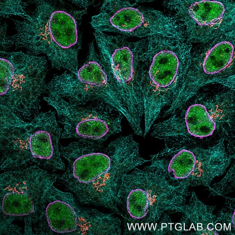
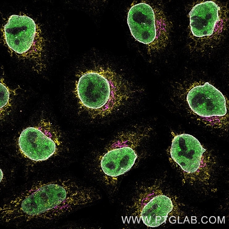
Immunofluorescence of Hela cells: PFA-fixed cells were stained with rabbit IgG anti-TDP43 antibody (10782-2-AP) labeled with FlexAble 2.0 CoraLite Plus 488 Kit (KFA501, green), rabbit IgG anti-CoxIV antibody (11242-1-AP) labeled with FlexAble 2.0 CoraLite Plus 555 Kit (KFA502, yellow), rabbit IgG anti-GM130 antibody (11308-1-AP) labeled with Multi-rAb CoraLite Plus 647 Goat anti-rabbit Secondary (RGAR005, magenta) and rabbit IgG anti-Lamin antibody (12987-1-AP) labeled with FlexAble 2.0 CoraLite Plus 750 Kit (KFA504, grey). Confocal images were acquired with a 63x oil objective and post-processed. Images were recorded at the Core Facility Bioimaging at the Biomedical Center, LMU Munich.
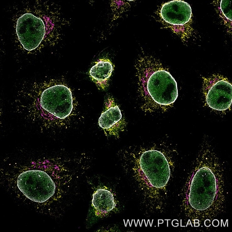
Immunofluorescence of Hela cells: PFA-fixed cells were stained with rabbit IgG anti-TDP43 antibody (10782-2-AP ) labeled with FlexAble 2.0 CoraLite Plus 488 Kit (KFA501, green), rabbit IgG anti-CoxIV antibody (11242-1-AP) labeled with FlexAble 2.0 CoraLite Plus 555 Kit (KFA502, yellow), rabbit IgG anti-GM130 antibody (11308-1-AP) labeled with Multi-rAb CoraLite Plus 647 Goat anti-rabbit Secondary (RGAR005, magenta) and rabbit IgG anti-Lamin antibody (12987-1-AP) labeled with FlexAble 2.0 CoraLite Plus 750 Kit (KFA504, grey).
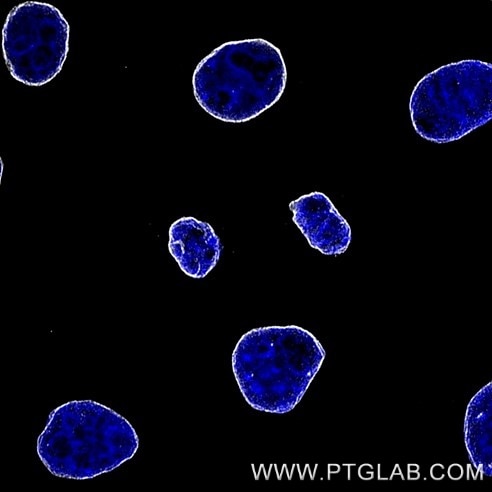
Immunofluorescence of Hela cells: PFA-fixed cells were stained with rabbit IgG anti-Lamin antibody (12987-1-AP) labeled with FlexAble 2.0 CoraLite Plus 750 Kit (KFA504, grey). Cell nuclei were stained with DAPI (blue). Confocal images were acquired with a 63x oil objective and post-processed. Images were recorded at the Core Facility Bioimaging at the Biomedical Center, LMU Munich.
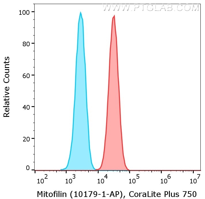
1X10^6 HeLa cells were intracellularly stained with anti-Mitofilin rabbit polyclonal antibody (10179-1-AP, red) or with isotype control antibody (30000-0-AP, blue) labeled with FlexAble 2.0 CoraLite Plus 750 Kit (KFA504). Cells were fixed and permeabilized with Foxp3 /Transcription Factor Staining Buffer Kit (PF00011).
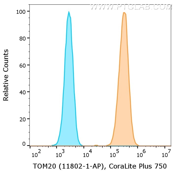
1X10^6 HeLa cells were intracellularly stained with anti-TOM20 rabbit polyclonal antibody (11802-1-AP, orange) or with isotype control antibody (30000-0-AP, blue) labeled with FlexAble 2.0 CoraLite Plus 750 Kit (KFA504). Cells were fixed and permeabilized with Foxp3 /Transcription Factor Staining Buffer Kit (PF00011).
|

|
|
Immunofluorescence of Hela cells: PFA-fixed cells were stained with rabbit IgG anti-Lamin antibody (12987-1-AP) labeled with FlexAble 2.0 CoraLite Plus 555 Kit (KFA502, yellow), rabbit IgG anti-GM130 antibody (11308-1-AP) labeled with FlexAble 2.0 CoraLite Plus 647 Kit (KFA503, magenta) and rabbit IgG anti-Tubulin antibody (80713-1-RR) labeled with FlexAble 2.0 CoraLite Plus 750 Kit (KFA504, grey). Cell nuclei were stained with DAPI (blue). Confocal images were acquired with a 63x oil objective and post-processed. Images were recorded at the Core Facility Bioimaging at the Biomedical Center, LMU Munich.
|
|
| 別包装 |
あり
|
| 種由来 |
Rabbit
|
| 交差種 |
Rabbit
|
| 適用 |
Western Blot
Immuno Fluorescence
Flow Cytometry
|
| 標識物 |
CoraLite(R) 750
|
| 励起波長 |
755
|
| 蛍光波長 |
780
|
|
| メーカー |
品番 |
包装 |
|
PGI
|
KFA504
|
10 RXN
|
※表示価格について
|
※当社では商品情報の適切な管理に努めておりますが、表示される法規制情報は最新でない可能性があります。
また法規制情報の表示が無いものは、必ずしも法規制に非該当であることを示すものではありません。
商品のお届け前に最新の製品法規制情報をお求めの際はこちらへお問い合わせください。
|
※当社取り扱いの試薬・機器製品および受託サービス・創薬支援サービス(納品物、解析データ等)は、研究用としてのみ販売しております。
人や動物の医療用・臨床診断用・食品用としては、使用しないように、十分ご注意ください。
法規制欄に体外診断用医薬品と記載のものは除きます。
|
|
※リンク先での文献等のダウンロードに際しましては、掲載元の規約遵守をお願いします。
|


