|
※サムネイル画像をクリックすると拡大画像が表示されます。
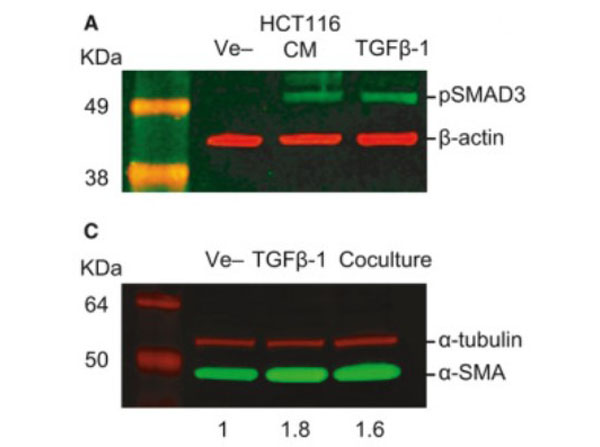
Western Blot using Sheep Anti-Rabbit IgG DyLight?800.TGF‐β‐mediated crosstalk between pericytes and CRC cells modulates pericyte secretome. (A) Incubation in HCT116 CM for 1?h induces SMAD3 phosphorylation in PC, as assessed by western blot. Exogenous recombinant TGF‐β (10?ng・mL−1) was used as a positive control, and β‐actin was used as loading control (n?=?3). (C) Western blot showing increased expression of αSMA in PC cocultured with HCT116 cells or stimulated with 10?ng・mL−1?TGF‐β1 for 48?h (n?=?3). α‐tubulin was used as loading control. Numbers indicate the expression fold change relative to the loading control. Fig. 5. PMID: 32767843.
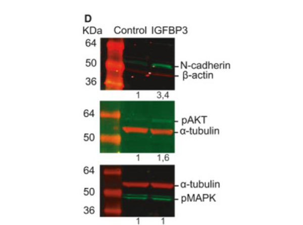
Western Blot results using Sheep Anti-Rabbit IgG DyLight?800.Insulin‐like growth factor‐binding protein 3 increases CRC cell migration and invasion through Akt activation. (D) Treatment with 50?ng・mL−1?IGFBP‐3 for 72?h promotes the expression of N‐cadherin in HCT116 cells as assessed by western blot (top panel). Phosphorylation status of Akt (middle panel) and MAPK (bottom panel) in HCT116 cells treated with 50?ng・mL−1?IGFBP‐3 for 15?min. Representative images of three independent experiments (n?=?3). Numbers indicate the expression fold change relative to the loading control. Fig. 7. PMID: 32767843.
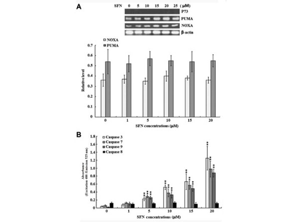
Western Blot results using Sheep Anti-Rabbit IgG DyLight?800.Effect of SFN on p73 expression and caspase enzyme activities. SW480 cells were treated with 0, 1, 5, 10, 15 and 20 μM SFN for 24 h. (A) The protein expression levels of p73, PUMA and NOXA were examined by western blotting. Quantified band densities are summarized below the images of the bands, and target protein expression levels were normalized to that of b-actin. (B) The activity of caspase-3, −7, −8 and −9 was determined using specific ELISA kits. The results of three independent experiments are shown. **P<0.01 vs. 0 μM SFN. SFN, sulforaphane; PUMA, p53 upregulated modulator of apoptosis; NOXA, phorbol-12-myristate-13-acetate-induced protein 1; ELISA, enzyme-linked immunosorbent assay. Figure?2. PMID: 28944886.
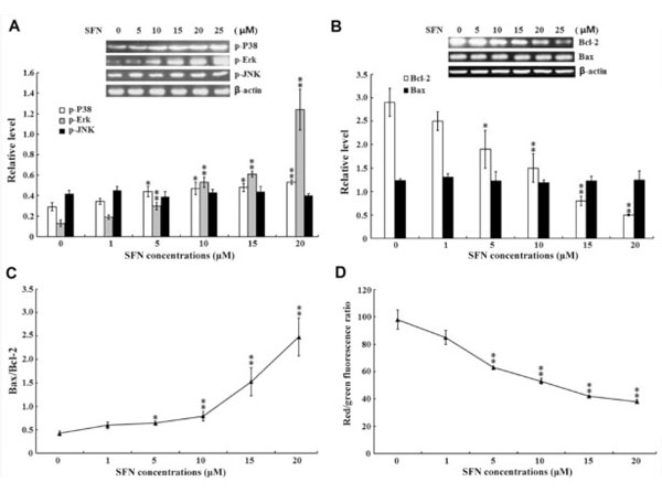
Western Blot results using Sheep Anti-Rabbit IgG DyLight?800.Effect of SFN on MAPKs and intrinsic apoptotic signaling pathways. SW480 cells were treated with 0, 1, 5, 10, 15 and 20 μM SFN for 24 h. (A) The protein expression levels of (A) p-p38, p-Erk and p-JNK and (B) Bcl-2 and Bax were examined by western blotting, and the quantified results are summarized below the images of the protein bands. Target protein expression levels were normalized to that of b-actin. (C) The Bax/Bcl-2 ratio. (D) The MMP was examined using a MMP assay kit with a JC-1 probe. The results of three independent experiments are shown. *P<0.05 and **P<0.01 vs. 0 μM SFN. SFN, sulforaphane; MAPK, mitogen-activated protein kinases; p-, phosphorylated; Erk, extracellular signal-regulated kinases; JNK, c-Jun N-terminal kinases; Bcl-2, B-cell lymphoma-2; Bax, Bcl-2-associated protein X; MMP, mitochondrial membrane potential. Figure?3. PMID: 28944886.
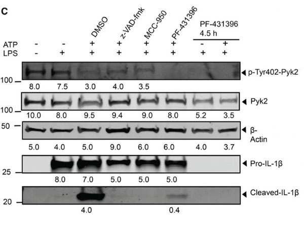
Western Blot Results using Sheep Anti-Rabbit IgG DyLight?800.LPL stabilizes Pyk2-ASC interaction by regulating Pyk2 localization.(C) WT BMDMs were primed with LPS and stimulated for NLRP3 activation by ATP (30 min) in the absence and presence of z-VAD-fmk (50 μM, 1 h), MCC-950 (50 μM, 1 h), and PF-431396 (25 μM) for 1 h or 4.5 h. Cell lysates were probed for Pyk2 phosphorylation and IL-1β products by immunoblotting. The density of each band is shown below. Here, β-actin is the total cell control. Figure 4. PMID: 35294888.
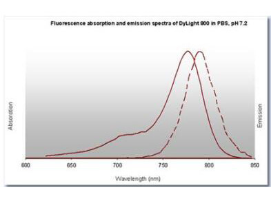
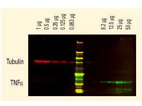
DyLight? dyes can be used for two-color western blot detection with low background and high signal.? Anti-tubulin was detected using a DyLight? 680 conjugate.? Anti-TNFa was detected using a DyLight? 800 conjugate. The image was captured using the OdysseyR Infrared Imaging System developed by LI-COR.
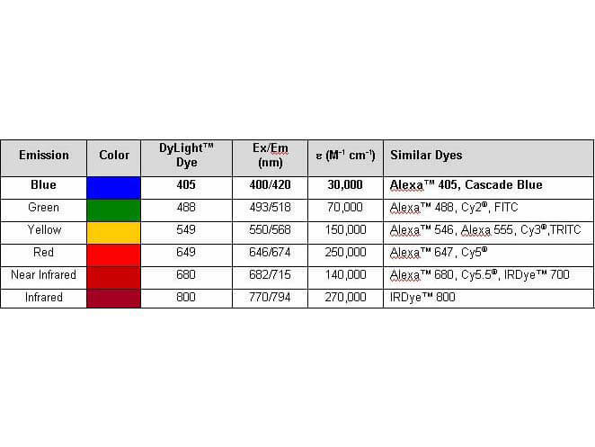
Properties of DyLight? Conjugates.
|
|
|
|
Western Blot using Sheep Anti-Rabbit IgG DyLight?800.TGF‐β‐mediated crosstalk between pericytes and CRC cells modulates pericyte secretome. (A) Incubation in HCT116 CM for 1?h induces SMAD3 phosphorylation in PC, as assessed by western blot. Exogenous recombinant TGF‐β (10?ng・mL−1) was used as a positive control, and β‐actin was used as loading control (n?=?3). (C) Western blot showing increased expression of αSMA in PC cocultured with HCT116 cells or stimulated with 10?ng・mL−1?TGF‐β1 for 48?h (n?=?3). α‐tubulin was used as loading control. Numbers indicate the expression fold change relative to the loading control. Fig. 5. PMID: 32767843.
|
|
| 別品名 |
Sheep Anti Rabbit IgG Antibody DyLight 800TM Conjugate, Sheep Anti-Rabbit IgG DyLight 800TM Conjugated Antibody
|
| 交差種 |
Rabbit
|
| 非交差(吸収処理)種 |
Human
Mouse
Rat
Bovine
Chicken
Sheep
Goat
Guinea Pig
Equine
|
| 免疫動物 |
Sheep
|
| 標識物 |
DyLightTM 800
|
| 精製度 |
Affinity Purified
|
| 参考文献 |
[Pub Med ID]28944886
|
| [注意事項] |
濃度はロットによって異なる可能性があります。メーカーDS及びCoAからご確認ください。
|
|
| メーカー |
品番 |
包装 |
|
RKL
|
611-645-122
|
100 UG
|
※表示価格について
| 当社在庫 |
なし
|
| 納期目安 |
約10日
|
| 法規制 |
毒
|
| 保存温度 |
4℃
|
|
※当社では商品情報の適切な管理に努めておりますが、表示される法規制情報は最新でない可能性があります。
また法規制情報の表示が無いものは、必ずしも法規制に非該当であることを示すものではありません。
商品のお届け前に最新の製品法規制情報をお求めの際はこちらへお問い合わせください。
|
※当社取り扱いの試薬・機器製品および受託サービス・創薬支援サービス(納品物、解析データ等)は、研究用としてのみ販売しております。
人や動物の医療用・臨床診断用・食品用としては、使用しないように、十分ご注意ください。
法規制欄に体外診断用医薬品と記載のものは除きます。
|
|
※リンク先での文献等のダウンロードに際しましては、掲載元の規約遵守をお願いします。
|
|
※CAS Registry Numbers have not been verified by CAS and may be inaccurate.
|