
| 標識物 |
DyLightTM 488
|
| 精製度 |
Affinity Purified
|
| 適用 |
Immuno Fluorescence
Flow Cytometry
Dot Blot
|
| 免疫動物 |
Goat
|
| 形状 |
凍結乾燥品
|
| 参考文献 |
[Pub Med ID]30247737
|
| [注意事項] |
濃度はロットによって異なる可能性があります。メーカーDS及びCoAからご確認ください。
|
|
※サムネイル画像をクリックすると拡大画像が表示されます。
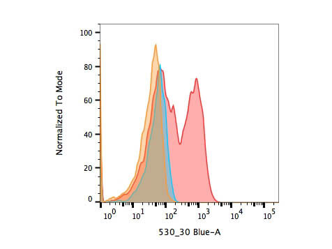
Flow Cytometry of Goat anti biotin DyLight 488 antibody. Cells were transiently transfected with a peroxidase encoding plasmid and were incubated for 24 hours before carrying out a modified TSA reaction. Cells were fixed and permeabilized before probing with Rockland goat anti biotin DyLight 488 at 1:5000 dilution for 2 hours at room temperature. Transfected and biotin supplemented population (red) is compared to transfected, non supplemented (blue) and non transfected, biotin supplemented (orange) controls. Image courtesy Ben Dyer, University of Pennsylvania.
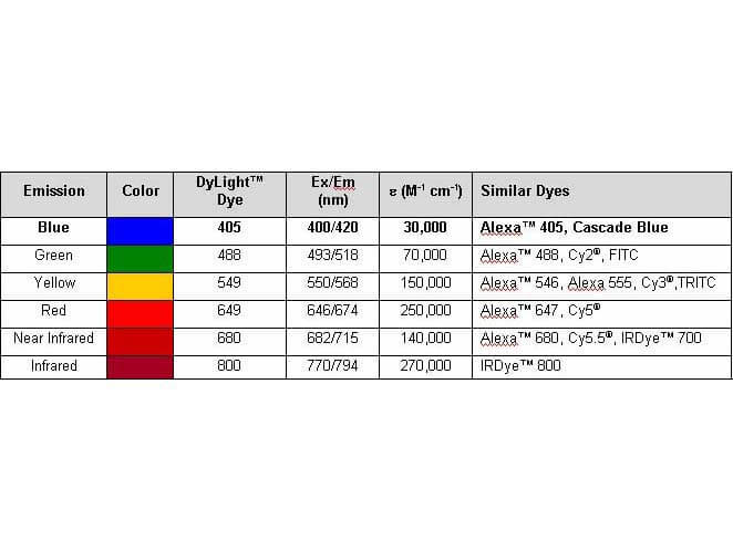
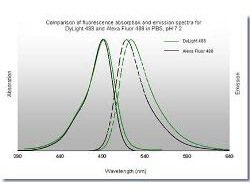
Properties of DyLightTM Fluorescent Dyes.
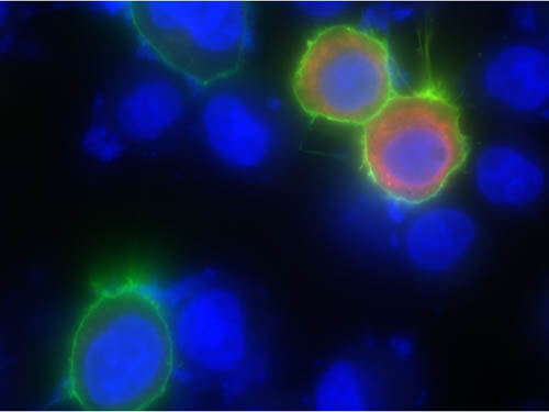
Immunofluorescence of Goat anti-biotin DyLight 488 antibody. Cells grown on glass coverslips were transiently transfected with a peroxidase-encoding plasmid and were incubated for 24 hours before carrying out a modified TSA reaction. Cells were fixed and permeabilized before probing with Rockland goat anti-biotin DyLight 488 at 1:1250 dilution for 2 hours at room temperature. Cells were washed before mounting on glass slides and imaged for biotin (green), DAPI (blue), and transfected peroxidase (red). Image courtesy Ben Dyer, University of Pennsylvania.
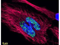
DyLightTM dyes can be used for multi-color immunofluorescence microscopy with uniform fluorescence intensity throughout the image. DyLightTM dyes are exceptionally bright and photostable and are optimized for microscopy and microarray detection methods. This image shows anti-histone detection using a DyLightTM 488 conjugate (green). Anti-Tubulin was detected using a DyLightTM 549 conjugate (red). Nuclei were counter-stained using DAPI (blue). The image was captured using an Axio Imager.Z1 (Zeiss Micro Imaging Inc).
Flow Cytometry of Goat anti-biotin DyLight 488 antibody. Cells were transiently transfected with a peroxidase-encoding plasmid and were incubated for 24 hours before carrying out a modified TSA reaction. Cells were fixed and permeabilized before probing with Rockland goat anti-biotin DyLight 488 at 1:5000 dilution for 2 hours at room temperature. Transfected and biotin supplemented population (red) is compared to transfected, non-supplemented (blue) and non-transfected, biotin supplemented (orange) controls. Image courtesy Ben Dyer, University of Pennsylvania.
|
|
|
|
Flow Cytometry of Goat anti biotin DyLight 488 antibody. Cells were transiently transfected with a peroxidase encoding plasmid and were incubated for 24 hours before carrying out a modified TSA reaction. Cells were fixed and permeabilized before probing with Rockland goat anti biotin DyLight 488 at 1:5000 dilution for 2 hours at room temperature. Transfected and biotin supplemented population (red) is compared to transfected, non supplemented (blue) and non transfected, biotin supplemented (orange) controls. Image courtesy Ben Dyer, University of Pennsylvania.
|
|
|
| メーカー |
品番 |
包装 |
|
RKL
|
600-141-098
|
100 UG
|
※表示価格について
| 当社在庫 |
なし
|
| 納期目安 |
約10日
|
| 法規制 |
毒
|
| 保存温度 |
4℃
|
|

