|
※サムネイル画像をクリックすると拡大画像が表示されます。
Figure 1. Western blot analysis of Galectin 1 using anti-Galectin 1 antibody (PB9240-1).
Electrophoresis was performed on a 5-20% SDS-PAGE gel at 70V (Stacking gel) / 90V (Resolving gel) for 2-3 hours. The sample well of each lane was loaded with 30 ug of sample under reducing conditions.
Lane 1: human Hela whole cell lysates,
Lane 2: human A549 whole cell lysates,
Lane 3: human A375 whole cell lysates,
Lane 4: human MCF-7 whole cell lysates,
Lane 5: rat kidney tissue lysates,
Lane 6: rat PC-12 whole cell lysates,
Lane 7: mouse kidney tissue lysates,
Lane 8: mouse NIH/3T3 whole cell lysates.
After electrophoresis, proteins were transferred to a nitrocellulose membrane at 150 mA for 50-90 minutes. Blocked the membrane with 5% non-fat milk/TBS for 1.5 hour at RT. The membrane was incubated with rabbit anti-Galectin 1 antigen affinity purified polyclonal antibody (Catalog # PB9240-1) at 0.5 μg/mL overnight at 4°C, then washed with TBS-0.1%Tween 3 times with 5 minutes each and probed with a goat anti-rabbit IgG-HRP secondary antibody at a dilution of 1:5000 for 1.5 hour at RT. The signal is developed using an Enhanced Chemiluminescent detection (ECL) kit (Catalog # EK1002) with Tanon 5200 system. A specific band was detected for Galectin 1 at approximately 15 kDa. The expected band size for Galectin 1 is at 15 kDa.

Figure 2. IHC analysis of Galectin 1 using anti-Galectin 1 antibody (PB9240-1).
Galectin 1 was detected in a paraffin-embedded section of mouse intestine cancer tissue. Heat mediated antigen retrieval was performed in EDTA buffer (pH 8.0, epitope retrieval solution). The tissue section was blocked with 10% goat serum. The tissue section was then incubated with 1 μg/ml rabbit anti-Galectin 1 Antibody (PB9240-1) overnight at 4°C. Biotinylated goat anti-rabbit IgG was used as secondary antibody and incubated for 30 minutes at 37°C. The tissue section was developed using Strepavidin-Biotin-Complex (SABC) (Catalog # SA1022) with DAB as the chromogen.

Figure 3. IHC analysis of Galectin 1 using anti-Galectin 1 antibody (PB9240-1).
Galectin 1 was detected in a paraffin-embedded section of rat kidney tissue. Heat mediated antigen retrieval was performed in EDTA buffer (pH 8.0, epitope retrieval solution). The tissue section was blocked with 10% goat serum. The tissue section was then incubated with 1 μg/ml rabbit anti-Galectin 1 Antibody (PB9240-1) overnight at 4°C. Biotinylated goat anti-rabbit IgG was used as secondary antibody and incubated for 30 minutes at 37°C. The tissue section was developed using Strepavidin-Biotin-Complex (SABC) (Catalog # SA1022) with DAB as the chromogen.
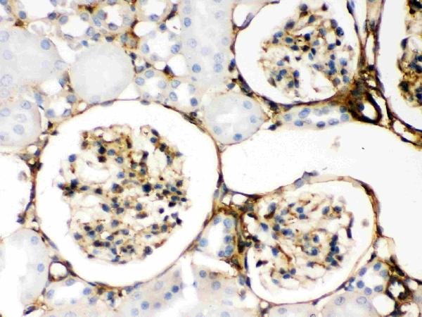
Figure 4. IHC analysis of Galectin 1 using anti-Galectin 1 antibody (PB9240-1).
Galectin 1 was detected in a paraffin-embedded section of human placenta tissue. Heat mediated antigen retrieval was performed in EDTA buffer (pH 8.0, epitope retrieval solution). The tissue section was blocked with 10% goat serum. The tissue section was then incubated with 1 μg/ml rabbit anti-Galectin 1 Antibody (PB9240-1) overnight at 4°C. Biotinylated goat anti-rabbit IgG was used as secondary antibody and incubated for 30 minutes at 37°C. The tissue section was developed using Strepavidin-Biotin-Complex (SABC) (Catalog # SA1022) with DAB as the chromogen.

Figure 5. IF analysis of Galectin 1 using anti-Galectin 1 antibody (PB9240-1).
Galectin 1 was detected in immunocytochemical section of U20S cells. Enzyme antigen retrieval was performed using IHC enzyme antigen retrieval reagent (AR0022) for 15 mins. The cells were blocked with 10% goat serum. And then incubated with 2μg/mL rabbit anti-Galectin 1 Antibody (PB9240-1) overnight at 4°C. DyLightR488 Conjugated Goat Anti-Rabbit IgG (BA1127) was used as secondary antibody at 1:100 dilution and incubated for 30 minutes at 37°C. The section was counterstained with DAPI. Visualize using a fluorescence microscope and filter sets appropriate for the label used.

Figure 6. Flow Cytometry analysis of PC-3 cells using anti-Galectin 1 antibody (PB9240-1).
Overlay histogram showing PC-3 cells stained with PB9240-1 (Blue line). To facilitate intracellular staining, cells were fixed with 4% paraformaldehyde and permeabilized with permeabilization buffer. The cells were blocked with 10% normal goat serum. And then incubated with rabbit anti-Galectin 1 Antibody (PB9240-1, 1μg/1x106 cells) for 30 min at 20°C. DyLightR488 conjugated goat anti-rabbit IgG (BA1127, 5-10μg/1x106 cells) was used as secondary antibody for 30 minutes at 20°C. Isotype control antibody (Green line) was rabbit IgG (1μg/1x106) used under the same conditions. Unlabelled sample without incubation with primary antibody and secondary antibody (Red line) was used as a blank control.

|
|
|
|
Figure 1. Western blot analysis of Galectin 1 using anti-Galectin 1 antibody (PB9240-1).
Electrophoresis was performed on a 5-20% SDS-PAGE gel at 70V (Stacking gel) / 90V (Resolving gel) for 2-3 hours. The sample well of each lane was loaded with 30 ug of sample under reducing conditions.
Lane 1: human Hela whole cell lysates,
Lane 2: human A549 whole cell lysates,
Lane 3: human A375 whole cell lysates,
Lane 4: human MCF-7 whole cell lysates,
Lane 5: rat kidney tissue lysates,
Lane 6: rat PC-12 whole cell lysates,
Lane 7: mouse kidney tissue lysates,
Lane 8: mouse NIH/3T3 whole cell lysates.
After electrophoresis, proteins were transferred to a nitrocellulose membrane at 150 mA for 50-90 minutes. Blocked the membrane with 5% non-fat milk/TBS for 1.5 hour at RT. The membrane was incubated with rabbit anti-Galectin 1 antigen affinity purified polyclonal antibody (Catalog # PB9240-1) at 0.5 μg/mL overnight at 4°C, then washed with TBS-0.1%Tween 3 times with 5 minutes each and probed with a goat anti-rabbit IgG-HRP secondary antibody at a dilution of 1:5000 for 1.5 hour at RT. The signal is developed using an Enhanced Chemiluminescent detection (ECL) kit (Catalog # EK1002) with Tanon 5200 system. A specific band was detected for Galectin 1 at approximately 15 kDa. The expected band size for Galectin 1 is at 15 kDa.
|
|
| 別品名 |
Galectin-1;Gal-1;14 kDa laminin-binding protein;HLBP14;14 kDa lectin;Beta-galactoside-binding lectin L-14-I;Galaptin;HBL;HPL;Lactose-binding lectin 1;Lectin galactoside-binding soluble 1;Putative MAPK-activating protein PM12;S-Lac lectin 1;LGALS1;
|
| 種由来 |
Human
|
| 交差種 |
Human
Mouse
Rat
|
| 適用 |
Western Blot
Enzyme Linked Immunosorbent Assay
Immunohistochemistry
Immuno Fluorescence
Immunocytochemistry (cell)
Flow Cytometry
|
| 免疫動物 |
Rabbit
|
| 抗体クラス |
IgG
|
| 標識物 |
Horseradish Peroxidase
|
| 精製度 |
Affinity Purified
|
| GENE ID |
3956
|
| Accession No.(Gene/Protein) |
P09382
|
| Gene Symbol |
LGALS1
|
| 分子量 |
14716 MW
|
| 概要 |
Boster Bio Anti-Galectin 1/LGALS1 Antibody Picoband® catalog # PB9240-1. Tested in ELISA, Flow Cytometry, IF, IHC, ICC, WB applications. This antibody reacts with Human, Mouse, Rat. The brand Picoband indicates this is a premium antibody that guarantees superior quality, high affinity, and strong signals with minimal background in Western blot applications. Only our best-performing antibodies are designated as Picoband, ensuring unmatched performance.
|
| 参考文献 |
1. Das, K., Lin, Y., Widen, E., Zhang, Y., Scherer, P. E. Chromosomal localization, expression pattern, and promoter analysis of the mouse gene encoding adipocyte-specific secretory protein Acrp30. Biochem. Biophys. Res. Commun. 280: 1120-1129, 2001.
2. Schaffler, A., Orso, E., Palitzsch, K.-D., Buchler, C., Drobnik, W., Furst, A., Scholmerich, J., Schmitz, G. The human apM-1, an adipocyte-specific gene linked to the family of TNF's and to genes expressed in activated T cells, is mapped to chromosome 1q21.3-q23, a susceptibility locus identified for familial combined hyperlipidaemia (FCH). Biochem. Biophys. Res. Commun. 260: 416-425, 1999.
|
|
| メーカー |
品番 |
包装 |
|
BBT
|
PB9240-1-HRP
|
100 UG
|
※表示価格について
| 当社在庫 |
なし
|
| 納期目安 |
1週間程度
|
| 保存温度 |
-20℃
|
|
