|
※サムネイル画像をクリックすると拡大画像が表示されます。
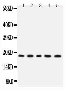
Figure 1. Western blot analysis of IL2 using anti-IL2 antibody (PB9013).
Electrophoresis was performed on a 5-20% SDS-PAGE gel at 70V (Stacking gel) / 90V (Resolving gel) for 2-3 hours. The sample well of each lane was loaded with 50ug of sample under reducing conditions.
Lane 1: Rat Thymus Tissue Lysate,
Lane 2: Rat Liver Tissue Lysate,
Lane 3: NRK Whole Cell Lysate,
Lane 4: PC12 Whole Cell Lysate,
Lane 5: RH35 Whole Cell Lysate.
After Electrophoresis, proteins were transferred to a Nitrocellulose membrane at 150mA for 50-90 minutes. Blocked the membrane with 5% Non-fat Milk/ TBS for 1.5 hour at RT. The membrane was incubated with rabbit anti-IL2 antigen affinity purified polyclonal antibody (Catalog # PB9013) at 0.5 μg/mL overnight at 4°C, then washed with TBS-0.1%Tween 3 times with 5 minutes each and probed with a goat anti-rabbit IgG-HRP secondary antibody at a dilution of 1:10000 for 1.5 hour at RT. The signal is developed using an Enhanced Chemiluminescent detection (ECL) kit (Catalog # EK1002) with Tanon 5200 system. A specific band was detected for IL2 at approximately 18KD. The expected band size for IL2 is at 18KD.
|
|
|
|
Figure 1. Western blot analysis of IL2 using anti-IL2 antibody (PB9013).
Electrophoresis was performed on a 5-20% SDS-PAGE gel at 70V (Stacking gel) / 90V (Resolving gel) for 2-3 hours. The sample well of each lane was loaded with 50ug of sample under reducing conditions.
Lane 1: Rat Thymus Tissue Lysate,
Lane 2: Rat Liver Tissue Lysate,
Lane 3: NRK Whole Cell Lysate,
Lane 4: PC12 Whole Cell Lysate,
Lane 5: RH35 Whole Cell Lysate.
After Electrophoresis, proteins were transferred to a Nitrocellulose membrane at 150mA for 50-90 minutes. Blocked the membrane with 5% Non-fat Milk/ TBS for 1.5 hour at RT. The membrane was incubated with rabbit anti-IL2 antigen affinity purified polyclonal antibody (Catalog # PB9013) at 0.5 μg/mL overnight at 4°C, then washed with TBS-0.1%Tween 3 times with 5 minutes each and probed with a goat anti-rabbit IgG-HRP secondary antibody at a dilution of 1:10000 for 1.5 hour at RT. The signal is developed using an Enhanced Chemiluminescent detection (ECL) kit (Catalog # EK1002) with Tanon 5200 system. A specific band was detected for IL2 at approximately 18KD. The expected band size for IL2 is at 18KD.
|
|
| 別品名 |
Interleukin-2;IL-2;T-cell growth factor;TCGF;Il2;Il-2;
|
| 種由来 |
Rat
|
| 交差種 |
Rat
|
| 免疫動物 |
Rabbit
|
| 抗体クラス |
IgG
|
| 標識物 |
Fluorescein Isothiocyanate
|
| 精製度 |
Affinity Purified
|
| GENE ID |
116562
|
| Accession No.(Gene/Protein) |
P17108
|
| Gene Symbol |
IL2
|
| 分子量 |
17633 MW
|
| 参考文献 |
1. Hollander, G. A.; Zuklys, S.; Morel, C.; Mizoguchi, E.; Mobisson, K.; Simpson, S.; Terhorst, C.; Wishart, W.; Golan, D. E.; Bhan, A. K.; Burakoff, S. J. Monoallelic expression of the interleukin-2 locus. Science 279: 2118-2121, 1998. 2. Lowenthal, J. W.; Zubler, R. H.; Nabholz, M.; MacDonald, H. R. Similarities between interleukin-2 receptor number and affinity on activated B and T lymphocytes. Nature 315: 669-672, 1985. 3. Shows, T.; Eddy, R.; Haley, L.; Byers, M.; Henry, M.; Fujita, T.; Matsui, H.; Taniguchi, T. Interleukin 2 (IL2) is assigned to human chromosome 4. Somat. Cell Molec. Genet. 10: 315-318, 1984.
|
|
| メーカー |
品番 |
包装 |
|
BBT
|
PB9013-FITC
|
100 UG
|
※表示価格について
| 当社在庫 |
なし
|
| 納期目安 |
1週間程度
|
| 保存温度 |
-20℃
|
|
※当社では商品情報の適切な管理に努めておりますが、表示される法規制情報は最新でない可能性があります。
また法規制情報の表示が無いものは、必ずしも法規制に非該当であることを示すものではありません。
商品のお届け前に最新の製品法規制情報をお求めの際はこちらへお問い合わせください。
|
※当社取り扱いの試薬・機器製品および受託サービス・創薬支援サービス(納品物、解析データ等)は、研究用としてのみ販売しております。
人や動物の医療用・臨床診断用・食品用としては、使用しないように、十分ご注意ください。
法規制欄に体外診断用医薬品と記載のものは除きます。
|
|
※リンク先での文献等のダウンロードに際しましては、掲載元の規約遵守をお願いします。
|
|
※CAS Registry Numbers have not been verified by CAS and may be inaccurate.
|


