|
※サムネイル画像をクリックすると拡大画像が表示されます。
Figure 1. Western blot analysis of ABI1 using anti-ABI1 antibody (PA1001).
Electrophoresis was performed on a 5-20% SDS-PAGE gel at 70V (Stacking gel) / 90V (Resolving gel) for 2-3 hours. The sample well of each lane was loaded with 30 ug of sample under reducing conditions.
Lane 1: human 293T whole cell lysates,
Lane 2: human MCF-7 whole cell lysates,
Lane 3: human U87 whole cell lysates,
Lane 4: rat brain tissue lysates,
Lane 5: rat C6 whole cell lysates,
Lane 6: mouse brain tissue lysates,
Lane 7: mouse Neuro-2a whole cell lysates,
Lane 8: mouse C2C12 whole cell lysates.
After electrophoresis, proteins were transferred to a nitrocellulose membrane at 150 mA for 50-90 minutes. Blocked the membrane with 5% non-fat milk/TBS for 1.5 hour at RT. The membrane was incubated with rabbit anti-ABI1 antigen affinity purified polyclonal antibody (Catalog # PA1001) at 0.5 μg/mL overnight at 4°C, then washed with TBS-0.1%Tween 3 times with 5 minutes each and probed with a goat anti-rabbit IgG-HRP secondary antibody at a dilution of 1:5000 for 1.5 hour at RT. The signal is developed using an Enhanced Chemiluminescent detection (ECL) kit (Catalog # EK1002) with Tanon 5200 system. A specific band was detected for ABI1 at approximately 60-65 kDa. The expected band size for ABI1 is at 55 kDa.

Figure 2. IHC analysis of ABI1 using anti-ABI1 antibody (PA1001).
ABI1 was detected in a paraffin-embedded section of human lung cancer tissue. Heat mediated antigen retrieval was performed in EDTA buffer (pH 8.0, epitope retrieval solution). The tissue section was blocked with 10% goat serum. The tissue section was then incubated with 2 μg/ml rabbit anti-ABI1 Antibody (PA1001) overnight at 4°C. Peroxidase Conjugated Goat Anti-rabbit IgG was used as secondary antibody and incubated for 30 minutes at 37°C. The tissue section was developed using HRP Conjugated Rabbit IgG Super Vision Assay Kit (Catalog # SV0002) with DAB as the chromogen.
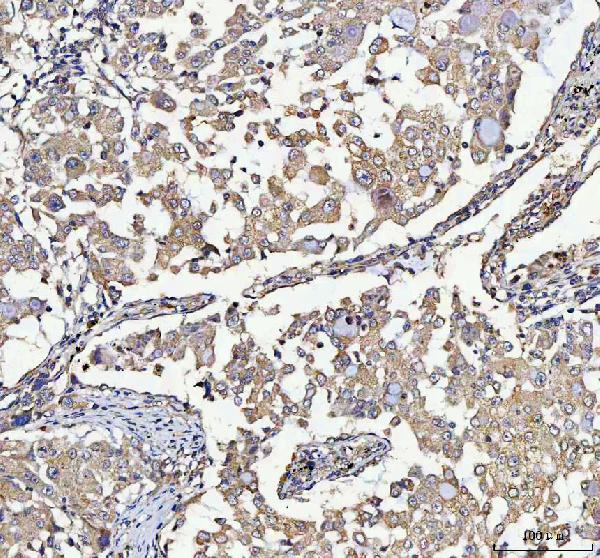
Figure 5. IF analysis of ABI1 using anti-ABI1 antibody (PA1001).
ABI1 was detected in a paraffin-embedded section of human intestine cancer tissue. Heat mediated antigen retrieval was performed in EDTA buffer (pH 8.0, epitope retrieval solution). The tissue section was blocked with 10% goat serum. The tissue section was then incubated with 5 μg/mL rabbit anti-ABI1 Antibody (PA1001) overnight at 4°C. Cy3 Conjugated Goat Anti-Rabbit IgG (BA1032) was used as secondary antibody at 1:500 dilution and incubated for 30 minutes at 37°C. The section was counterstained with DAPI. Visualize using a fluorescence microscope and filter sets appropriate for the label used.

Figure 3. IHC analysis of ABI1 using anti-ABI1 antibody (PA1001).
ABI1 was detected in a paraffin-embedded section of human colorectal adenocarcinoma tissue. Heat mediated antigen retrieval was performed in EDTA buffer (pH 8.0, epitope retrieval solution). The tissue section was blocked with 10% goat serum. The tissue section was then incubated with 2 μg/ml rabbit anti-ABI1 Antibody (PA1001) overnight at 4°C. Peroxidase Conjugated Goat Anti-rabbit IgG was used as secondary antibody and incubated for 30 minutes at 37°C. The tissue section was developed using HRP Conjugated Rabbit IgG Super Vision Assay Kit (Catalog # SV0002) with DAB as the chromogen.

Figure 6. IF analysis of ABI1 using anti-ABI1 antibody (PA1001).
ABI1 was detected in a paraffin-embedded section of human breast cancer tissue. Heat mediated antigen retrieval was performed in EDTA buffer (pH 8.0, epitope retrieval solution). The tissue section was blocked with 10% goat serum. The tissue section was then incubated with 5 μg/mL rabbit anti-ABI1 Antibody (PA1001) overnight at 4°C. Cy3 Conjugated Goat Anti-Rabbit IgG (BA1032) was used as secondary antibody at 1:500 dilution and incubated for 30 minutes at 37°C. The section was counterstained with DAPI. Visualize using a fluorescence microscope and filter sets appropriate for the label used.

Figure 4. IF analysis of ABI1 using anti-ABI1 antibody (PA1001).
ABI1 was detected in an immunocytochemical section of U2OS cells. Enzyme antigen retrieval was performed using IHC enzyme antigen retrieval reagent (AR0022) for 15 mins. The cells were blocked with 10% goat serum. And then incubated with 5 μg/mL rabbit anti-ABI1 Antibody (PA1001) overnight at 4°C. Cy3 Conjugated Goat Anti-Rabbit IgG (BA1032) was used as secondary antibody at 1:500 dilution and incubated for 30 minutes at 37°C. The section was counterstained with DAPI. Visualize using a fluorescence microscope and filter sets appropriate for the label used.
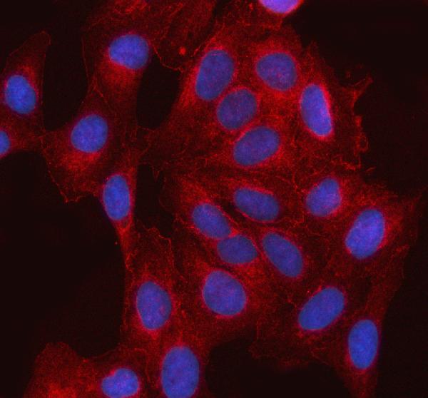
Figure 7. Flow Cytometry analysis of MCF-7 cells using anti-ABI1 antibody (PA1001).
Overlay histogram showing MCF-7 cells stained with PA1001 (Blue line). To facilitate intracellular staining, cells were fixed with 4% paraformaldehyde and permeabilized with permeabilization buffer. The cells were blocked with 10% normal goat serum. And then incubated with rabbit anti-ABI1 Antibody (PA1001, 1 μg/1x106 cells) for 30 min at 20°C. DyLightR488 conjugated goat anti-rabbit IgG (BA1127, 5-10 μg/1x106 cells) was used as secondary antibody for 30 minutes at 20°C. Isotype control antibody (Green line) was rabbit IgG (1 μg/1x106) used under the same conditions. Unlabelled sample (Red line) was also used as a control.

|
|
|
|
Figure 1. Western blot analysis of ABI1 using anti-ABI1 antibody (PA1001).
Electrophoresis was performed on a 5-20% SDS-PAGE gel at 70V (Stacking gel) / 90V (Resolving gel) for 2-3 hours. The sample well of each lane was loaded with 30 ug of sample under reducing conditions.
Lane 1: human 293T whole cell lysates,
Lane 2: human MCF-7 whole cell lysates,
Lane 3: human U87 whole cell lysates,
Lane 4: rat brain tissue lysates,
Lane 5: rat C6 whole cell lysates,
Lane 6: mouse brain tissue lysates,
Lane 7: mouse Neuro-2a whole cell lysates,
Lane 8: mouse C2C12 whole cell lysates.
After electrophoresis, proteins were transferred to a nitrocellulose membrane at 150 mA for 50-90 minutes. Blocked the membrane with 5% non-fat milk/TBS for 1.5 hour at RT. The membrane was incubated with rabbit anti-ABI1 antigen affinity purified polyclonal antibody (Catalog # PA1001) at 0.5 μg/mL overnight at 4°C, then washed with TBS-0.1%Tween 3 times with 5 minutes each and probed with a goat anti-rabbit IgG-HRP secondary antibody at a dilution of 1:5000 for 1.5 hour at RT. The signal is developed using an Enhanced Chemiluminescent detection (ECL) kit (Catalog # EK1002) with Tanon 5200 system. A specific band was detected for ABI1 at approximately 60-65 kDa. The expected band size for ABI1 is at 55 kDa.
|
|
| 別品名 |
Abl interactor 1;Abelson interactor 1;Abi-1;Abl-binding protein 4;AblBP4;Eps8 SH3 domain-binding protein;Eps8-binding protein;Nap1-binding protein;Nap1BP;Spectrin SH3 domain-binding protein 1;e3B1;ABI1;SSH3BP1;
|
| 種由来 |
Human
|
| 交差種 |
Human
Mouse
Rat
|
| 適用 |
Western Blot
Immunohistochemistry
Immuno Fluorescence
Immunocytochemistry (cell)
Flow Cytometry
|
| 免疫動物 |
Rabbit
|
| 抗体クラス |
IgG
|
| 標識物 |
Cyanine 3
|
| 精製度 |
Affinity Purified
|
| Accession No.(Gene/Protein) |
Q8IZP0
|
| Gene Symbol |
ABI1
|
| 分子量 |
55081 MW
|
| 概要 |
Boster Bio Anti-SSH3BP1/ABI1 Antibody catalog # PA1001. Tested in Flow Cytometry, IF, IHC, ICC, WB applications. This antibody reacts with Human, Mouse, Rat. The brand Picoband indicates this is a premium antibody that guarantees superior quality, high affinity, and strong signals with minimal background in Western blot applications. Only our best-performing antibodies are designated as Picoband, ensuring unmatched performance.
|
| 参考文献 |
1. Tani, K.; Sato, S.; Sukezane, T.; Kojima, H.; Hirose, H.; Hanafusa, H.; Shishido, T. : Abl interactor 1 promotes tyrosine 296 phosphorylation of mammalian Enabled (Mena) by c-Abl kinase. J. Biol. Chem. 278: 21685-21692, 2003.
2. Taki, T.; Shibuya, N.; Taniwaki, M.; Hanada, R.; Morishita, K.; Bessho, F.; Yanagisawa, M.; Hayashi, Y. : ABI-1, a human homolog to mouse Abl-interactor 1, fuses the MLL gene in acute myeloid leukemia with t(10;11)(p11.2;q23). Blood 92: 1125-1130, 1998.
|
|
| メーカー |
品番 |
包装 |
|
BBT
|
PA1001-CY3
|
100 UG
|
※表示価格について
| 当社在庫 |
なし
|
| 納期目安 |
1週間程度
|
| 保存温度 |
-20℃
|
|
