
| 別品名 |
14-3-3 protein zeta/delta; Protein kinase C inhibitor protein 1; KCIP-1; YWHAZ
|
| 種由来 |
Human
|
| 標識物 |
DyLightTM 488
|
| 精製度 |
Affinity Purified
|
| 適用 |
Western Blot
Flow Cytometry
|
| 免疫動物 |
Mouse
|
| クローン |
6G5
|
| 交差種 |
Human
Mouse
Rat
Monkey
|
| GENE ID |
7534
|
| Accession No.(Gene/Protein) |
P63104
|
| Gene Symbol |
YWHAZ
|
| 感度 |
>5000 cells
|
| 形状 |
凍結乾燥品
|
| 参考文献 |
1. "Entrez Gene: YWHAZ tyrosine 3-monooxygenase/tryptophan 5-monooxygenase activation protein, zeta polypeptide".
2. Nishimura Y, Komatsu S, Ichikawa D, Nagata H, Hirajima S, Takeshita H, Kawaguchi T, Arita T, Konishi H, Kashimoto K, Shiozaki A, Fujiwara H, Okamoto K, Tsuda H, Otsuji E (Apr 2013). "Overexpression of YWHAZ relates to tumor cell proliferation and malignant outcome of gastric carcinoma". British Journal of Cancer. 108 (6): 1324?31.
|
|
※サムネイル画像をクリックすると拡大画像が表示されます。
Figure 1. Western blot analysis of 14-3-3 zeta/delta using anti-14-3-3 zeta/delta antibody (M01141).
Electrophoresis was performed on a 5-20% SDS-PAGE gel at 70V (Stacking gel) / 90V (Resolving gel) for 2-3 hours. The sample well of each lane was loaded with 50ug of sample under reducing conditions.
Lane 1: human Hela whole cell lysates;
Lane 2: human A549 whole cell lysates;
Lane 3: monkey COS-7 whole cell lysates;
Lane 4: human Raji whole cell lysates;
Lane 5:huamn Caco-2 whole cell lysates;
Lane 6: huamn Jurkat whole cell lysates;
Lane 7: mouse brain tissue lysates;
Lane 8: rat brain tissue lysates
After Electrophoresis, proteins were transferred to a Nitrocellulose membrane at 150mA for 50-90 minutes. Blocked the membrane with 5% Non-fat Milk/ TBS for 1.5 hour at RT. The membrane was incubated with mouse anti-14-3-3 zeta/delta antigen affinity purified monoclonal antibody (Catalog # M01141) at 0.5 μg/mL overnight at 4°C, then washed with TBS-0.1%Tween 3 times with 5 minutes each and probed with a goat anti-mouse IgG-HRP secondary antibody at a dilution of 1:10000 for 1.5 hour at RT. The signal is developed using an Enhanced Chemiluminescent detection (ECL) kit (Catalog # EK1001) with Tanon 5200 system. A specific band was detected for 14-3-3 zeta/delta at approximately 28KD. The expected band size for 14-3-3 zeta/delta is at 28KD.
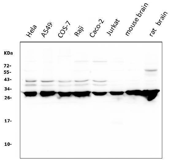
Figure 2. Flow Cytometry analysis of PC-3 cells using anti-14-3-3 zeta/delta antibody (M01141).Overlay histogram showing PC-3 cells stained with M01141 (Blue line). To facilitate intracellular staining, cells were fixed with 4% paraformaldehyde and permeabilized with permeabilization buffer. The cells were blocked with 10% normal goat serum. And then incubated with mouse anti-14-3-3 zeta/delta Antibody (M01141,1μg/1x106 cells) for 30 min at 20°C. DyLight?488 conjugated goat anti-mouse IgG (BA1126, 5-10μg/1x106 cells) was used as secondary antibody for 30 minutes at 20°C. Isotype control antibody (Green line) was mouse IgG (1μg/1x106) used under the same conditions. Unlabelled sample without incubation with primary antibody and secondary antibody (Red line) was used as a blank control.
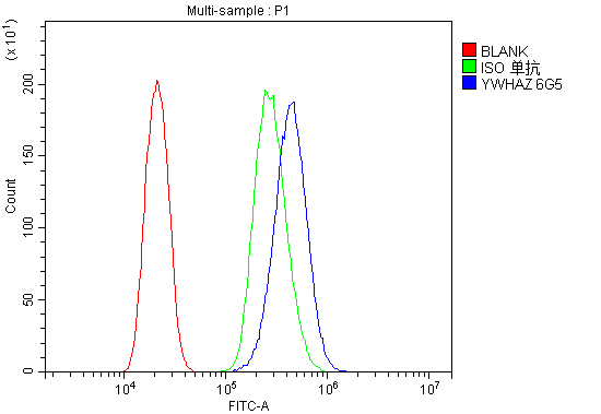
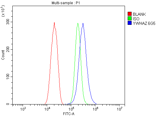
Figure 3. Flow Cytometry analysis of SiHa cells using anti-14-3-3 zeta/delta antibody (M01141).Overlay histogram showing SiHa cells stained with M01141 (Blue line). To facilitate intracellular staining, cells were fixed with 4% paraformaldehyde and permeabilized with permeabilization buffer. The cells were blocked with 10% normal goat serum. And then incubated with mouse anti-14-3-3 zeta/delta Antibody (M01141,1μg/1x106 cells) for 30 min at 20°C. DyLight?488 conjugated goat anti-mouse IgG (BA1126, 5-10μg/1x106 cells) was used as secondary antibody for 30 minutes at 20°C. Isotype control antibody (Green line) was mouse IgG (1μg/1x106) used under the same conditions. Unlabelled sample without incubation with primary antibody and secondary antibody (Red line) was used as a blank control.
|
|
|
|
Figure 1. Western blot analysis of 14-3-3 zeta/delta using anti-14-3-3 zeta/delta antibody (M01141).
Electrophoresis was performed on a 5-20% SDS-PAGE gel at 70V (Stacking gel) / 90V (Resolving gel) for 2-3 hours. The sample well of each lane was loaded with 50ug of sample under reducing conditions.
Lane 1: human Hela whole cell lysates;
Lane 2: human A549 whole cell lysates;
Lane 3: monkey COS-7 whole cell lysates;
Lane 4: human Raji whole cell lysates;
Lane 5:huamn Caco-2 whole cell lysates;
Lane 6: huamn Jurkat whole cell lysates;
Lane 7: mouse brain tissue lysates;
Lane 8: rat brain tissue lysates
After Electrophoresis, proteins were transferred to a Nitrocellulose membrane at 150mA for 50-90 minutes. Blocked the membrane with 5% Non-fat Milk/ TBS for 1.5 hour at RT. The membrane was incubated with mouse anti-14-3-3 zeta/delta antigen affinity purified monoclonal antibody (Catalog # M01141) at 0.5 μg/mL overnight at 4°C, then washed with TBS-0.1%Tween 3 times with 5 minutes each and probed with a goat anti-mouse IgG-HRP secondary antibody at a dilution of 1:10000 for 1.5 hour at RT. The signal is developed using an Enhanced Chemiluminescent detection (ECL) kit (Catalog # EK1001) with Tanon 5200 system. A specific band was detected for 14-3-3 zeta/delta at approximately 28KD. The expected band size for 14-3-3 zeta/delta is at 28KD.
|
|
|
| メーカー |
品番 |
包装 |
|
BBT
|
M01141-DYLIGHT488
|
100 UG
|
※表示価格について
| 当社在庫 |
なし
|
| 納期目安 |
1週間程度
|
| 保存温度 |
-20℃
|
|

