
| 別品名 |
High mobility group protein B1; High mobility group protein 1; HMG-1; HMGB1; HMG1
|
| 種由来 |
Human
|
| 標識物 |
DyLightTM 594
|
| 適用 |
Western Blot
Immunohistochemistry
Flow Cytometry
|
| 免疫動物 |
Mouse
|
| クローン |
5H3
|
| 交差種 |
Human
Mouse
Rat
Monkey
|
| GENE ID |
3146
|
| Accession No.(Gene/Protein) |
P09429
|
| Gene Symbol |
HMGB1
|
| 形状 |
凍結乾燥品
|
| 参考文献 |
1. Ferrari S, Finelli P, Rocchi M, Bianchi ME (July 1996). "The active gene that encodes human high mobility group 1 protein (HMG1) contains introns and maps to chromosome 13".?Genomics.?35?(2): 367?71.
2. Klune JR, Dhupar R, Cardinal J, Billiar TR, Tsung A (2008)."HMGB1: endogenous danger signaling".?Mol. Med.?14?(7-8): 476?84.
3. Yang H, Hreggvidsdottir HS, Palmblad K, Wang H, Ochani M, Li J, Lu B, Chavan S, Rosas-Ballina M, Al-Abed Y, Akira S, Bierhaus A, Erlandsson-Harris H, Andersson U, Tracey KJ (June 2010).?"A critical cysteine is required for HMGB1 binding to Toll-like receptor 4 and activation of macrophage cytokine release".?Proc. Natl. Acad. Sci. U.S.A.?107?(26): 11942?7.
|
|
※サムネイル画像をクリックすると拡大画像が表示されます。
Figure 1. Western blot analysis of HMGB1 using anti-HMGB1 antibody (M00066-2).
Electrophoresis was performed on a 5-20% SDS-PAGE gel at 70V (Stacking gel) / 90V (Resolving gel) for 2-3 hours. The sample well of each lane was loaded with 50ug of sample under reducing conditions.
Lane 1: human HepG2 whole cell lysates
Lane 2: human CCRF-CEM whole cell lysates
Lane 3: monkey COS-7 whole cell lysates
Lane 4: human SW620 whole cell lysates
Lane 5: human THP-1 whole cell lysates
Lane 6: rat PC-12 whole cell lysates
Lane 7: rat RH35 whole cell lysates
Lane 8: mouse NIH/3T3 whole cell lysates
After Electrophoresis, proteins were transferred to a Nitrocellulose membrane at 150mA for 50-90 minutes. Blocked the membrane with 5% Non-fat Milk/ TBS for 1.5 hour at RT. The membrane was incubated with mouse anti-HMGB1 antigen affinity purified monoclonal antibody (Catalog # M00066-2) at 0.5 μg/mL overnight at 4°C, then washed with TBS-0.1%Tween 3 times with 5 minutes each and probed with a goat anti-mouse IgG-HRP secondary antibody at a dilution of 1:10000 for 1.5 hour at RT. The signal is developed using an Enhanced Chemiluminescent detection (ECL) kit (Catalog # EK1001) with Tanon 5200 system. A specific band was detected for HMGB1 at approximately 25KD. The expected band size for HMGB1 is at 25KD.

Figure 10. IHC analysis of HMGB1 using anti-HMGB1 antibody (M00066-2). HMGB1 was detected in paraffin-embedded section of rat brain tissues. Heat mediated antigen retrieval was performed in citrate buffer (pH6, epitope retrieval solution) for 20 mins. The tissue section was blocked with 10% goat serum. The tissue section was then incubated with 1μg/ml mouse anti-HMGB1 Antibody (M00066-2) overnight at 4°C. Biotinylated goat anti-mouse IgG was used as secondary antibody and incubated for 30 minutes at 37°C. The tissue section was developed using Strepavidin-Biotin-Complex (SABC)(Catalog # SA1021) with DAB as the chromogen.
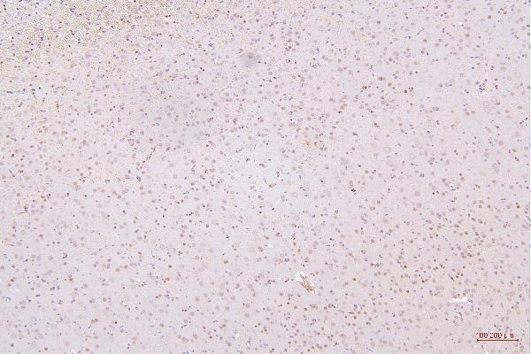
Figure 2. IHC analysis of HMGB1 using anti-HMGB1 antibody (M00066-2).
HMGB1 was detected in paraffin-embedded section of human intestinal cancer tissues. Heat mediated antigen retrieval was performed in citrate buffer (pH6, epitope retrieval solution) for 20 mins. The tissue section was blocked with 10% goat serum. The tissue section was then incubated with 1μg/ml mouse anti-HMGB1 Antibody (M00066-2) overnight at 4°C. Biotinylated goat anti-mouse IgG was used as secondary antibody and incubated for 30 minutes at 37°C. The tissue section was developed using Strepavidin-Biotin-Complex (SABC)(Catalog # SA1021) with DAB as the chromogen.

Figure 3. IHC analysis of HMGB1 using anti-HMGB1 antibody (M00066-2).
HMGB1 was detected in paraffin-embedded section of human intestinal cancer tissues. Heat mediated antigen retrieval was performed in citrate buffer (pH6, epitope retrieval solution) for 20 mins. The tissue section was blocked with 10% goat serum. The tissue section was then incubated with 1μg/ml mouse anti-HMGB1 Antibody (M00066-2) overnight at 4°C. Biotinylated goat anti-mouse IgG was used as secondary antibody and incubated for 30 minutes at 37°C. The tissue section was developed using Strepavidin-Biotin-Complex (SABC)(Catalog # SA1021) with DAB as the chromogen.

Figure 4. IHC analysis of HMGB1 using anti-HMGB1 antibody (M00066-2).
HMGB1 was detected in paraffin-embedded section of human intestinal cancer tissues. Heat mediated antigen retrieval was performed in citrate buffer (pH6, epitope retrieval solution) for 20 mins. The tissue section was blocked with 10% goat serum. The tissue section was then incubated with 1μg/ml mouse anti-HMGB1 Antibody (M00066-2) overnight at 4°C. Biotinylated goat anti-mouse IgG was used as secondary antibody and incubated for 30 minutes at 37°C. The tissue section was developed using Strepavidin-Biotin-Complex (SABC)(Catalog # SA1021) with DAB as the chromogen.

Figure 5. IHC analysis of HMGB1 using anti-HMGB1 antibody (M00066-2).
HMGB1 was detected in paraffin-embedded section of human mammary cancer tissues. Heat mediated antigen retrieval was performed in citrate buffer (pH6, epitope retrieval solution) for 20 mins. The tissue section was blocked with 10% goat serum. The tissue section was then incubated with 1μg/ml mouse anti-HMGB1 Antibody (M00066-2) overnight at 4°C. Biotinylated goat anti-mouse IgG was used as secondary antibody and incubated for 30 minutes at 37°C. The tissue section was developed using Strepavidin-Biotin-Complex (SABC)(Catalog # SA1021) with DAB as the chromogen.

Figure 6. IHC analysis of HMGB1 using anti-HMGB1 antibody (M00066-2).
HMGB1 was detected in paraffin-embedded section of human mammary cancer tissues. Heat mediated antigen retrieval was performed in citrate buffer (pH6, epitope retrieval solution) for 20 mins. The tissue section was blocked with 10% goat serum. The tissue section was then incubated with 1μg/ml mouse anti-HMGB1 Antibody (M00066-2) overnight at 4°C. Biotinylated goat anti-mouse IgG was used as secondary antibody and incubated for 30 minutes at 37°C. The tissue section was developed using Strepavidin-Biotin-Complex (SABC)(Catalog # SA1021) with DAB as the chromogen.

Figure 7. IHC analysis of HMGB1 using anti-HMGB1 antibody (M00066-2).
HMGB1 was detected in paraffin-embedded section of human lung cancer tissues. Heat mediated antigen retrieval was performed in citrate buffer (pH6, epitope retrieval solution) for 20 mins. The tissue section was blocked with 10% goat serum. The tissue section was then incubated with 1μg/ml mouse anti-HMGB1 Antibody (M00066-2) overnight at 4°C. Biotinylated goat anti-mouse IgG was used as secondary antibody and incubated for 30 minutes at 37°C. The tissue section was developed using Strepavidin-Biotin-Complex (SABC)(Catalog # SA1021) with DAB as the chromogen.

Figure 8. IHC analysis of HMGB1 using anti-HMGB1 antibody (M00066-2).
HMGB1 was detected in paraffin-embedded section of human lung cancer tissues. Heat mediated antigen retrieval was performed in citrate buffer (pH6, epitope retrieval solution) for 20 mins. The tissue section was blocked with 10% goat serum. The tissue section was then incubated with 1μg/ml mouse anti-HMGB1 Antibody (M00066-2) overnight at 4°C. Biotinylated goat anti-mouse IgG was used as secondary antibody and incubated for 30 minutes at 37°C. The tissue section was developed using Strepavidin-Biotin-Complex (SABC)(Catalog # SA1021) with DAB as the chromogen.

Figure 9. IHC analysis of HMGB1 using anti-HMGB1 antibody (M00066-2).
HMGB1 was detected in paraffin-embedded section of rat brain tissues. Heat mediated antigen retrieval was performed in citrate buffer (pH6, epitope retrieval solution) for 20 mins. The tissue section was blocked with 10% goat serum. The tissue section was then incubated with 1μg/ml mouse anti-HMGB1 Antibody (M00066-2) overnight at 4°C. Biotinylated goat anti-mouse IgG was used as secondary antibody and incubated for 30 minutes at 37°C. The tissue section was developed using Strepavidin-Biotin-Complex (SABC)(Catalog # SA1021) with DAB as the chromogen.

|
|
|
|
Figure 1. Western blot analysis of HMGB1 using anti-HMGB1 antibody (M00066-2).
Electrophoresis was performed on a 5-20% SDS-PAGE gel at 70V (Stacking gel) / 90V (Resolving gel) for 2-3 hours. The sample well of each lane was loaded with 50ug of sample under reducing conditions.
Lane 1: human HepG2 whole cell lysates
Lane 2: human CCRF-CEM whole cell lysates
Lane 3: monkey COS-7 whole cell lysates
Lane 4: human SW620 whole cell lysates
Lane 5: human THP-1 whole cell lysates
Lane 6: rat PC-12 whole cell lysates
Lane 7: rat RH35 whole cell lysates
Lane 8: mouse NIH/3T3 whole cell lysates
After Electrophoresis, proteins were transferred to a Nitrocellulose membrane at 150mA for 50-90 minutes. Blocked the membrane with 5% Non-fat Milk/ TBS for 1.5 hour at RT. The membrane was incubated with mouse anti-HMGB1 antigen affinity purified monoclonal antibody (Catalog # M00066-2) at 0.5 μg/mL overnight at 4°C, then washed with TBS-0.1%Tween 3 times with 5 minutes each and probed with a goat anti-mouse IgG-HRP secondary antibody at a dilution of 1:10000 for 1.5 hour at RT. The signal is developed using an Enhanced Chemiluminescent detection (ECL) kit (Catalog # EK1001) with Tanon 5200 system. A specific band was detected for HMGB1 at approximately 25KD. The expected band size for HMGB1 is at 25KD.
|
|
|
| メーカー |
品番 |
包装 |
|
BBT
|
M00066-2-DYLIGHT594
|
100 UG
|
※表示価格について
| 当社在庫 |
なし
|
| 納期目安 |
1週間程度
|
| 保存温度 |
-20℃
|
|

