| 別品名 |
Solute carrier family 2, facilitated glucose transporter member 9; Glucose transporter type 9; GLUT-9; Urate transporter; SLC2A9; GLUT9
|
| 抗原部位 |
C-terminus
|
| 種由来 |
Human
|
| 標識物 |
Horseradish Peroxidase
|
| 精製度 |
Affinity Purified
|
| 適用 |
Western Blot
|
| 免疫動物 |
Rabbit
|
| 交差種 |
Human
Mouse
Rat
|
| GENE ID |
10215
|
| Accession No.(Gene/Protein) |
Q13516
|
| Gene Symbol |
OLIG2
|
| 形状 |
凍結乾燥品
|
| 参考文献 |
1. Buffo, A., Vosko, M. R., Erturk, D., Hamann, G. F., Jucker, M., Rowitch, D., Gotz, M. Expression pattern of the transcription factor Olig2 in response to brain injuries: implications for neuronal repair. Proc. Nat. Acad. Sci. 102: 18183-18188, 2005.
2. Georgieva, L., Moskvina, V., Peirce, T., Norton, N., Bray, N. J., Jones, L., Holmans, P., MacGregor, S., Zammit, S., Wilkinson, J., Williams, H., Nikolov, I., and 9 others. Convergent evidence that oligodendrocyte lineage transcription factor 2 (OLIG2) and interacting genes influence susceptibility to schizophrenia. Proc. Nat. Acad. Sci. 103: 12469-12474, 2006.
3. Huang, K., Tang, W., Tang, R., Xu, Z., He, Z., Li, Z., Xu, Y., Li, X., He, G., Feng, G., He, L., Shi, Y. Positive association between OLIG2 and schizophrenia in the Chinese Han population. Hum. Genet. 122: 659-660, 2008.
|
|
※サムネイル画像をクリックすると拡大画像が表示されます。
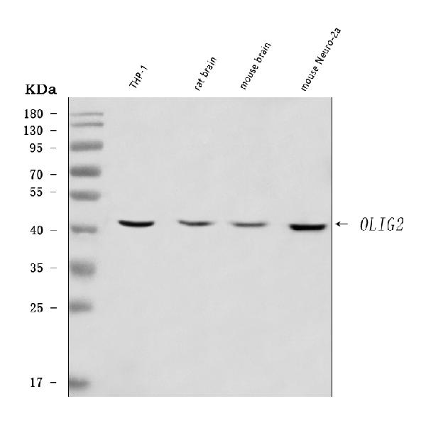
Figure 1. Western blot analysis of OLIG2 using anti-OLIG2 antibody (A02247-2).
Electrophoresis was performed on a 5-20% SDS-PAGE gel at 70V (Stacking gel) / 90V (Resolving gel) for 2-3 hours. The sample well of each lane was loaded with 30 ug of sample under reducing conditions.
Lane 1: human THP-1 whole cell lysates,
Lane 2: rat brain tissue lysates,
Lane 3: mouse brain tissue lysates,
Lane 4: mouse Neuro-2a whole cell lysates.
After electrophoresis, proteins were transferred to a nitrocellulose membrane at 150 mA for 50-90 minutes. Blocked the membrane with 5% non-fat milk/TBS for 1.5 hour at RT. The membrane was incubated with rabbit anti-OLIG2 antigen affinity purified polyclonal antibody (Catalog # A02247-2) at 0.5 μg/mL overnight at 4°C, then washed with TBS-0.1%Tween 3 times with 5 minutes each and probed with a goat anti-rabbit IgG-HRP secondary antibody at a dilution of 1:5000 for 1.5 hour at RT. The signal is developed using an Enhanced Chemiluminescent detection (ECL) kit (Catalog # EK1002) with Tanon 5200 system. A specific band was detected for OLIG2 at approximately 40 kDa. The expected band size for OLIG2 is at 32 kDa.

|

|
|
Figure 1. Western blot analysis of OLIG2 using anti-OLIG2 antibody (A02247-2).
Electrophoresis was performed on a 5-20% SDS-PAGE gel at 70V (Stacking gel) / 90V (Resolving gel) for 2-3 hours. The sample well of each lane was loaded with 30 ug of sample under reducing conditions.
Lane 1: human THP-1 whole cell lysates,
Lane 2: rat brain tissue lysates,
Lane 3: mouse brain tissue lysates,
Lane 4: mouse Neuro-2a whole cell lysates.
After electrophoresis, proteins were transferred to a nitrocellulose membrane at 150 mA for 50-90 minutes. Blocked the membrane with 5% non-fat milk/TBS for 1.5 hour at RT. The membrane was incubated with rabbit anti-OLIG2 antigen affinity purified polyclonal antibody (Catalog # A02247-2) at 0.5 μg/mL overnight at 4°C, then washed with TBS-0.1%Tween 3 times with 5 minutes each and probed with a goat anti-rabbit IgG-HRP secondary antibody at a dilution of 1:5000 for 1.5 hour at RT. The signal is developed using an Enhanced Chemiluminescent detection (ECL) kit (Catalog # EK1002) with Tanon 5200 system. A specific band was detected for OLIG2 at approximately 40 kDa. The expected band size for OLIG2 is at 32 kDa.
|
|
|
| メーカー |
品番 |
包装 |
|
BBT
|
A02247-2-HRP
|
100 UG
|
※表示価格について
| 当社在庫 |
なし
|
| 納期目安 |
1週間程度
|
| 保存温度 |
-20℃
|
|


