
| 別品名 |
Toll-like receptor 2;Toll/interleukin-1 receptor-like protein 4;CD282;TLR2;TIL4;
|
| 抗原部位 |
a.a.20-683
|
| 種由来 |
Mouse
|
| 標識物 |
Biotin
|
| 精製度 |
Affinity Purified
|
| 適用 |
Western Blot
Enzyme Linked Immunosorbent Assay
|
| 免疫動物 |
Rabbit
|
| 交差種 |
Mouse
|
| GENE ID |
24088
|
| Accession No.(Gene/Protein) |
Q9QUN7
|
| Gene Symbol |
Tlr2
|
| 分子量 |
89838 MW
|
| 形状 |
凍結乾燥品
|
| 参考文献 |
1. Aderem, A., Ulevitch, R. J. Toll-like receptors in the induction of the innate immune response. Nature 406: 782-785, 2000.
2. Alexopoulou, L., Thomas, V., Schnare, M., Lobet, Y., Anguita, J., Schoen, R. T., Medzhitov, R., Fikrig, E., Flavell, R. A. Hyporesponsiveness to vaccination with Borrelia burgdorferi OspA in humans and in TLR1- and TLR2-deficient mice. Nature Med. 8: 878-884, 2002.
3. Aliprantis, A. O., Yang, R.-B., Mark, M. R., Suggett, S., Devaux, B., Radolf, J. D., Klimpel, G. R., Godowski, P., Zychlinsky, A. Cell activation and apoptosis by bacterial lipoproteins through toll-like receptor-2. Science 285: 736-739, 1999.
|
|
※サムネイル画像をクリックすると拡大画像が表示されます。
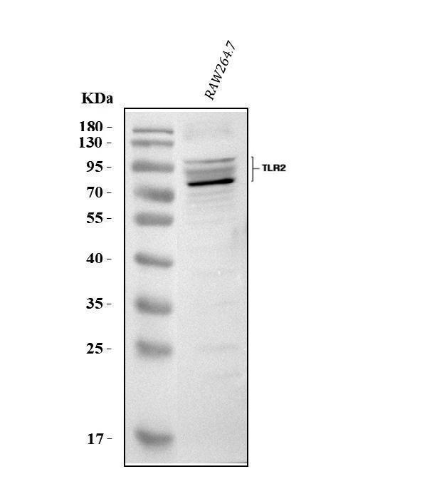
Figure 1. Western blot analysis of Tlr2 using anti-Tlr2 antibody (A00131-4).
Electrophoresis was performed on a 5-20% SDS-PAGE gel at 70V (Stacking gel) / 90V (Resolving gel) for 2-3 hours. The sample well of each lane was loaded with 30 ug of sample under reducing conditions.
Lane 1: mouse RAW264.7 whole cell lysates.
After electrophoresis, proteins were transferred to a nitrocellulose membrane at 150 mA for 50-90 minutes. Blocked the membrane with 5% non-fat milk/TBS for 1.5 hour at RT. The membrane was incubated with rabbit anti-Tlr2 antigen affinity purified polyclonal antibody (Catalog # A00131-4) at 0.5 μg/mL overnight at 4°C, then washed with TBS-0.1%Tween 3 times with 5 minutes each and probed with a goat anti-rabbit IgG-HRP secondary antibody at a dilution of 1:5000 for 1.5 hour at RT. The signal is developed using an Enhanced Chemiluminescent detection (ECL) kit (Catalog # EK1002) with Tanon 5200 system. A specific band was detected for Tlr2 at approximately 89 kDa. The expected band size for Tlr2 is at 89 kDa.
|
|
|
|
Figure 1. Western blot analysis of Tlr2 using anti-Tlr2 antibody (A00131-4).
Electrophoresis was performed on a 5-20% SDS-PAGE gel at 70V (Stacking gel) / 90V (Resolving gel) for 2-3 hours. The sample well of each lane was loaded with 30 ug of sample under reducing conditions.
Lane 1: mouse RAW264.7 whole cell lysates.
After electrophoresis, proteins were transferred to a nitrocellulose membrane at 150 mA for 50-90 minutes. Blocked the membrane with 5% non-fat milk/TBS for 1.5 hour at RT. The membrane was incubated with rabbit anti-Tlr2 antigen affinity purified polyclonal antibody (Catalog # A00131-4) at 0.5 μg/mL overnight at 4°C, then washed with TBS-0.1%Tween 3 times with 5 minutes each and probed with a goat anti-rabbit IgG-HRP secondary antibody at a dilution of 1:5000 for 1.5 hour at RT. The signal is developed using an Enhanced Chemiluminescent detection (ECL) kit (Catalog # EK1002) with Tanon 5200 system. A specific band was detected for Tlr2 at approximately 89 kDa. The expected band size for Tlr2 is at 89 kDa.
|
|
|
| メーカー |
品番 |
包装 |
|
BBT
|
A00131-4-BIOTIN
|
100 UG
|
※表示価格について
| 当社在庫 |
なし
|
| 納期目安 |
1週間程度
|
| 保存温度 |
-20℃
|
|

