|
※サムネイル画像をクリックすると拡大画像が表示されます。
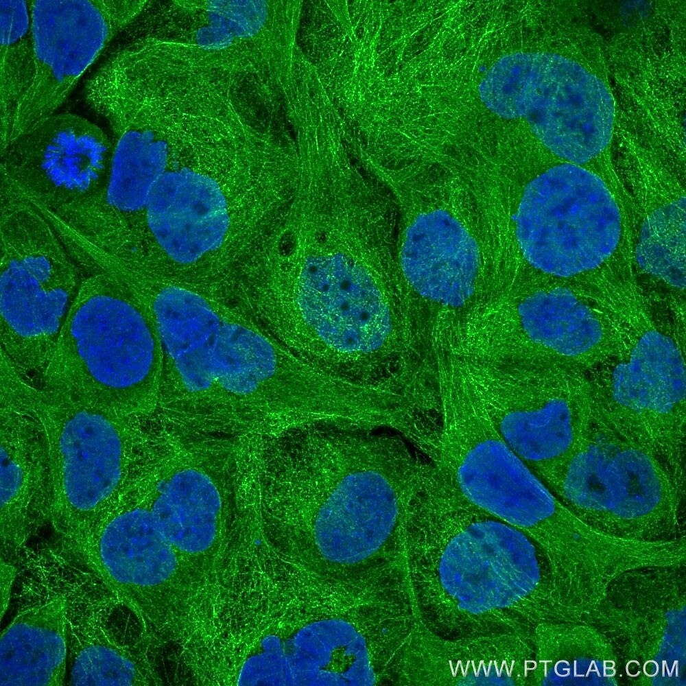
Immunofluorescence of A431 cells: PFA-fixed cells were stained with rat anti-Tubulin alpha antibody labeled with FlexAble CoraLite Plus 488 Kit (KFA121, green). Cell nuclei were stained with DAPI (blue). Confocal images were acquired with a 63x oil objective and post-processed. Images were recorded at the Core Facility Bioimaging at the Biomedical Center, LMU Munich.
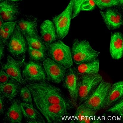
Immunofluorescence of A431: Formaldehyde-fixed A431 cells were stained with rat IgG2a kappa anti-Tubulin antibody labeled with FlexAble CoraLite Plus 488 Kit (KFA121, green) and rat IgG1 kappa anti-RPA32 antibody labeled with FlexAble CoraLite Plus 555 Kit (KFA122, red). Epifluorescence images were acquired with a 20x objective and post-processed.
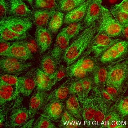
Immunofluorescence of A431: Formaldehyde-fixed A431 cells were stained with rat IgG2a kappa anti-Tubulin antibody labeled with FlexAble CoraLite Plus 488 Kit (KFA121, green) and rat IgG2b kappa anti-RNA PolymeraseII antibody labeled with FlexAble CoraLite Plus 555 Kit (KFA122, red). Epifluorescence images were acquired with a 20x objective and post-processed.
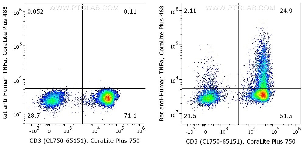
Human peripheral blood lymphocytes (PBMC) were either un-stimulated (left) or stimulated with PMA and Ionomycin for 6 hours in the presence of monensin (right). ?Cells were fixed and permeabilized with Foxp3/Transcription Factor Staining Buffer Kit (PF00011). After wash, cells were stained with rat anti-human TNF-α antibody labeled with FlexAble CoraLite Plus 488 for Rat Kappa Light Chain (KFA121) and CD3 antibody (CL750-65151).
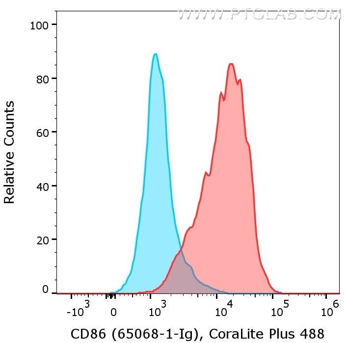
1X10^6 of LPS treated mouse splenocytes were surface stained with rat anti-Mouse CD86 (65068-1-Ig, Clone: GL1, red) or rat IgG2a isotype control (65209-1-Ig, blue) labeled with FlexAble CoraLite Plus 488 for Rat Kappa Light Chain (KFA121). Cells were not fixed.
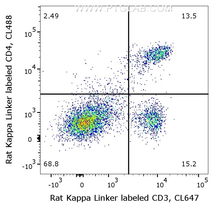
1X10^6 C57BL/6 mouse splenocytes were surface stained with 0.5μg rat anti-Mouse CD4 (65104-1-Ig, Clone: GK1.5) labeled with FlexAble CoraLite Plus 488 Kit (KFA121) and 0.5μg rat anti-Mouse CD3 (65077-1-Ig, Clone: 17A2) labeled with FlexAble CoraLite Plus 647 Kit (KFA123). Cells were not fixed.
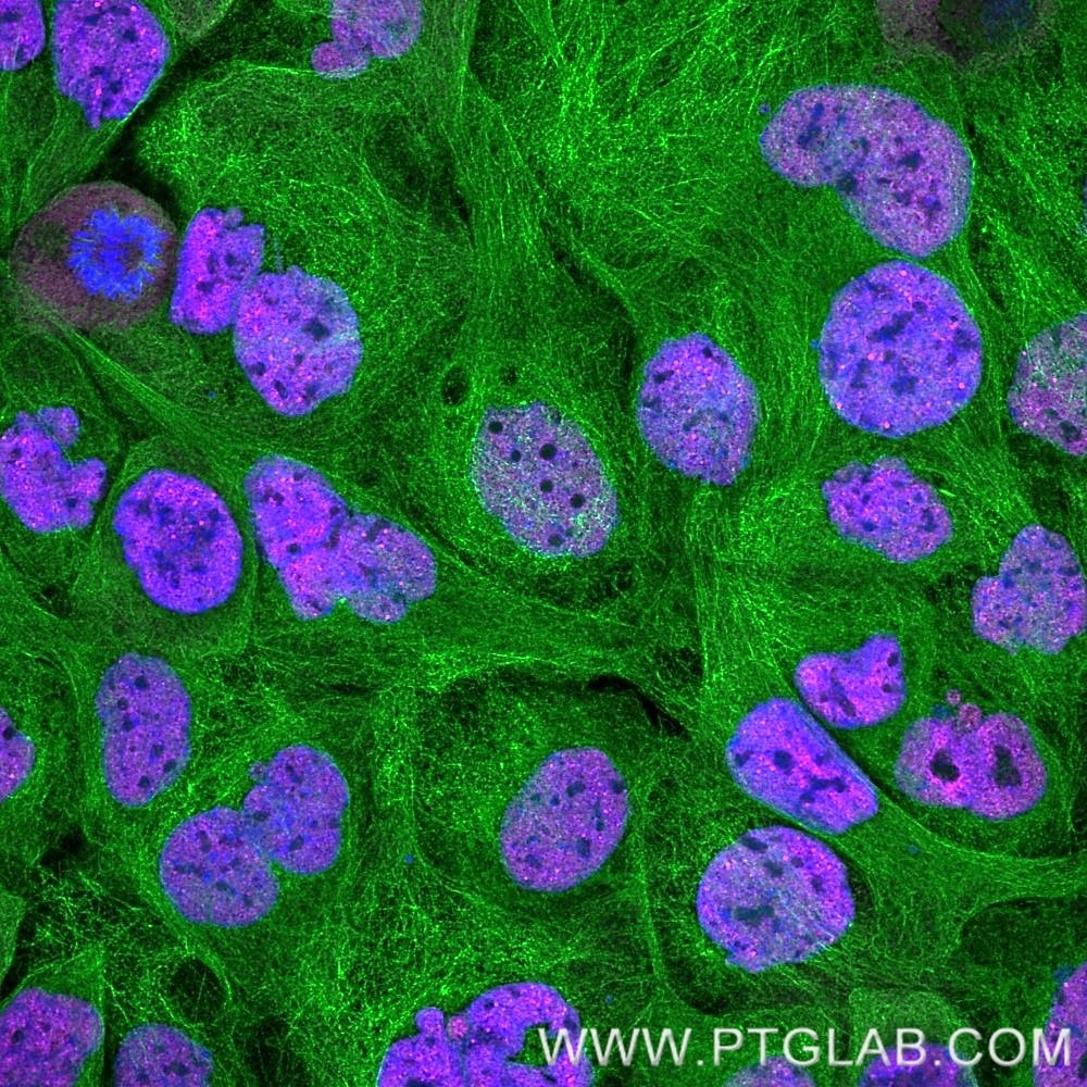
Immunofluorescence of A431 cells: PFA-fixed cells were co-stained with rat anti-RNA Pol II Ser5 antibody labeled with FlexAble CoraLite Plus 647 Kit (KFA123, magenta) and with rat anti-Tubulin alpha antibody labeled with FlexAble CoraLite Plus 488 Kit (KFA121, green). Cell nuclei were stained with DAPI (blue). Confocal images were acquired with a 63x oil objective and post-processed. Images were recorded at the Core Facility Bioimaging at the Biomedical Center, LMU Munich.
|

|
|
Immunofluorescence of A431 cells: PFA-fixed cells were stained with rat anti-Tubulin alpha antibody labeled with FlexAble CoraLite Plus 488 Kit (KFA121, green). Cell nuclei were stained with DAPI (blue). Confocal images were acquired with a 63x oil objective and post-processed. Images were recorded at the Core Facility Bioimaging at the Biomedical Center, LMU Munich.
|
|
| 別包装 |
あり
|
| 種由来 |
Rat
|
| 交差種 |
Rat
|
| 適用 |
Western Blot
Immuno Fluorescence
Flow Cytometry
|
| 標識物 |
CoraLite(R) 488
|
| 励起波長 |
493
|
| 蛍光波長 |
522
|
|
| メーカー |
品番 |
包装 |
|
PGI
|
KFA121
|
50 RXN
|
※表示価格について
| 販売状況 |
品番変更、サイズ変更
|
| 当社在庫 |
あり
|
| 法規制 |
安
|
| 保存温度 |
-20℃
|
|
※当社では商品情報の適切な管理に努めておりますが、表示される法規制情報は最新でない可能性があります。
また法規制情報の表示が無いものは、必ずしも法規制に非該当であることを示すものではありません。
商品のお届け前に最新の製品法規制情報をお求めの際はこちらへお問い合わせください。
|
※当社取り扱いの試薬・機器製品および受託サービス・創薬支援サービス(納品物、解析データ等)は、研究用としてのみ販売しております。
人や動物の医療用・臨床診断用・食品用としては、使用しないように、十分ご注意ください。
法規制欄に体外診断用医薬品と記載のものは除きます。
|
|
※リンク先での文献等のダウンロードに際しましては、掲載元の規約遵守をお願いします。
|


