|
※サムネイル画像をクリックすると拡大画像が表示されます。
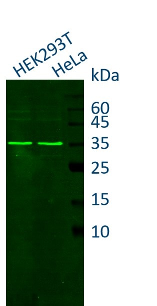
HEK-293 and Hela cell lysates were subjected to SDS-PAGE followed by fluorescent western blot analysis with rabbit anti-GAPDH antiboy (10494-1-AP) and Nano-Secondary alpaca anti-rabbit IgG, recombinant VHH, CoraLite Plus 488 (srb2GCL488-1), 1:500.

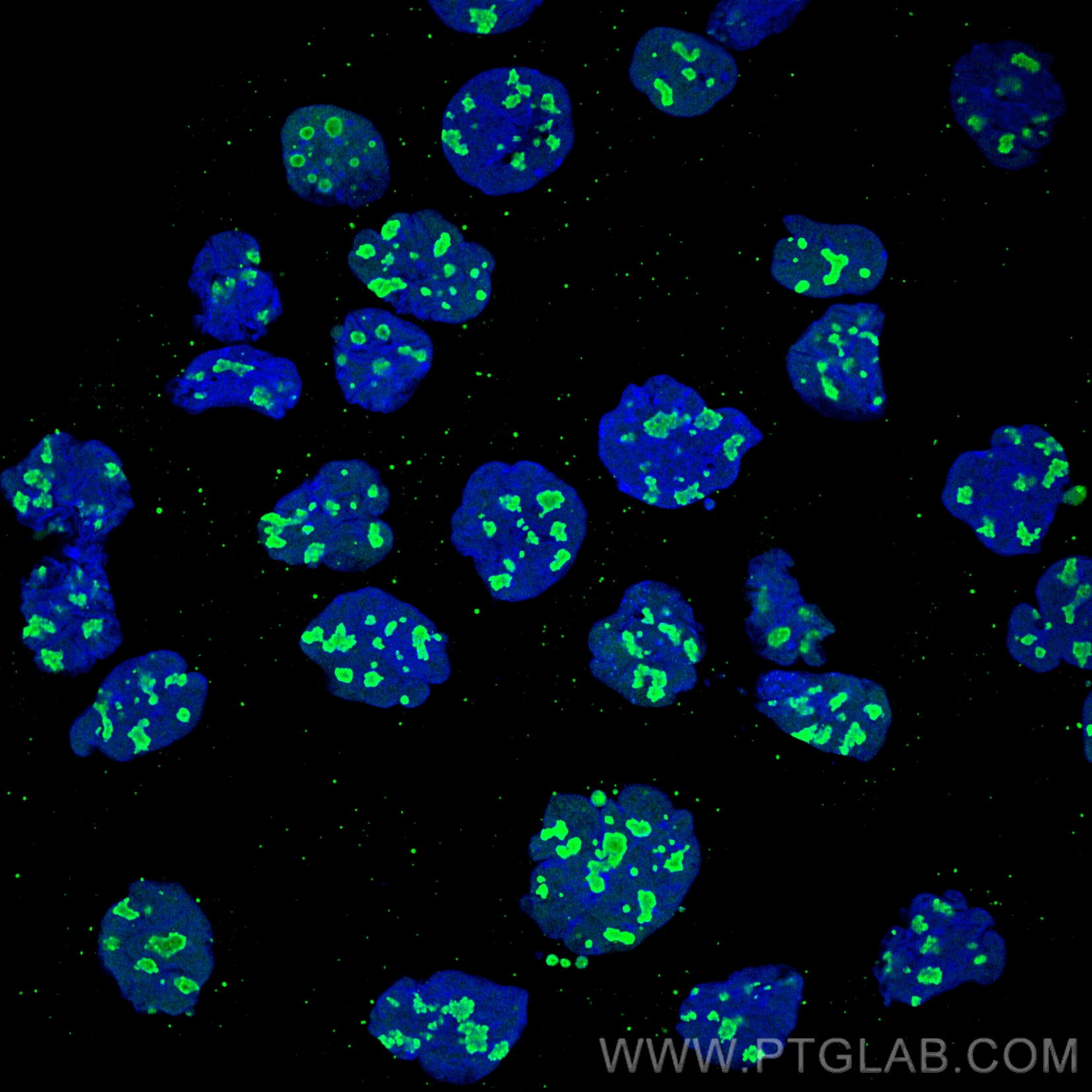
Immunofluorescence analysis of A431 cells stained with rabbit anti-NCL antibody (10556-1-AP, green) and Nano-Secondary alpaca anti-rabbit IgG, recombinant VHH, CoraLite Plus 488 (srb2GCL488-1), 1:500. Nuclei were stained with DAPI (blue). Images were recorded at the Core Facility Bioimaging at the Biomedical Center, LMU Munich.

Immunofluorescence analysis of A431 cells co-stained with anti-EGFR antibody (cetuximab biosimilar, huIgG1, magenta) and anti-NCL antibody (10556-1-AP, green). Nano-Secondary alpaca anti-human IgG, recombinant VHH, CoraLite Plus 647 (shuGCL647-2), 1:500 and Nano-Secondary alpaca anti-rabbit IgG, recombinant VHH, CoraLite Plus 488 (srb2GCL488-1), 1:500 were used for detection. Nuclei were stained with DAPI. Merged image on the right shows an overlay of both channels and DAPI (blue). ?Images were recorded at the Core Facility Bioimaging at the Biomedical Center, LMU Munich.

|

|
|
HEK-293 and Hela cell lysates were subjected to SDS-PAGE followed by fluorescent western blot analysis with rabbit anti-GAPDH antiboy (10494-1-AP) and Nano-Secondary alpaca anti-rabbit IgG, recombinant VHH, CoraLite Plus 488 (srb2GCL488-1), 1:500.
|
|
| 別品名 |
Alpaca single domain antibody, VHH, Nanobody, binding domain of single domain antibody, Nano-antibody
|
| 別包装 |
あり
|
| 種由来 |
Rabbit
|
| 交差種 |
Rabbit
|
| 非交差(吸収処理)種 |
Human
Mouse
Rat
|
| 適用 |
Western Blot
Immuno Fluorescence
|
| 免疫動物 |
Alpaca
|
| クローン |
CTK0119
CTK0120
|
| 標識物 |
CoraLite(R) 488
|
| その他 |
[備考]旧Chromotek(クロモテック、ドイツ)社商品。2020年10月よりProteintech Group(プロテインテック、米国)社ブランドの一部になりました。
|
|
| メーカー |
品番 |
包装 |
|
PGI
|
SRB2GCL488-1-100
|
100 UL
|
※表示価格について
|
※当社では商品情報の適切な管理に努めておりますが、表示される法規制情報は最新でない可能性があります。
また法規制情報の表示が無いものは、必ずしも法規制に非該当であることを示すものではありません。
商品のお届け前に最新の製品法規制情報をお求めの際はこちらへお問い合わせください。
|
※当社取り扱いの試薬・機器製品および受託サービス・創薬支援サービス(納品物、解析データ等)は、研究用としてのみ販売しております。
人や動物の医療用・臨床診断用・食品用としては、使用しないように、十分ご注意ください。
法規制欄に体外診断用医薬品と記載のものは除きます。
|
|
※リンク先での文献等のダウンロードに際しましては、掲載元の規約遵守をお願いします。
|
|
※CAS Registry Numbers have not been verified by CAS and may be inaccurate.
|


