|
※サムネイル画像をクリックすると拡大画像が表示されます。
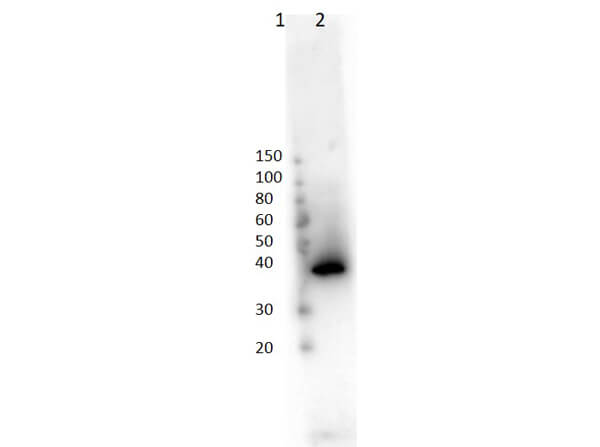
Western Blot of Rabbit Anti-Lac I Antibody. Lane 1: Molecular Weight Marker (3μL). Lane 2: recombinant LAC-I (0.05μg). Primary Antibody: Anti-LAC-I at 1:1000 for 1hr at RT. Secondary Antibody: Goat anti-Rabbit IgG HRP (p/n 611-103-122) at 1:40,000 for 30mins at RT. Block: BlockOut Buffer (p/n MB-073) for 30mins at RT. Predicted/Observed MW: ~38kDa.
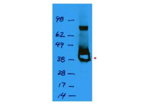
Western blot using Rockland's affinity purified anti-Lac I antibody shows detection of a 38 kDa band corresponding to recombinant Lac I (arrowhead). The blot was blocked with 5% BLOTTO (p/n B501-0500) in TTBS overnight at 4°C. Primary antibody was used at a 1:1,000 dilution followed by reaction with a 1:10,000 dilution of HRP goat anti-rabbit IgG. ECL was used for detection. Personal communication, S. Hughes & M. Abram, NCI, Bethesda, MD.
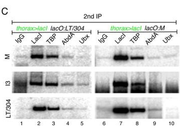
(C)?32P PCRs of cgChIPs from?thorax>lacI; lacO:LT/304?(left) and?thorax>lacI; lacO:M?(right) embryos. These PCRs assess the presence of?M, I3, and?LT/304?sequences following a secondary IP using the antibodies indicated above the gels (IgG, anti-LacI, anti-TBP, anti-AbdA, and anti-Ubx). The results confirm that IPs for LacI and TBP, but not abdominal Hox proteins, pull-down multiple?Dll?5’ regions in?Dll-expressing cells in the thorax. Fig 3. PMID:?22523743
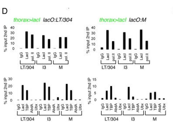
(D) qPCR measurements of cgChIP experiments for chromatin isolated from?thorax>lacI; lacO:LT/304?(left) and?thorax>lacI; lacO:M?(right). Measurements are for the three?Dll?sequences (LT/304, I3, and?M) after secondary IPs with the antibodies indicated (top gels: IgG, anti-LacI, anti-PolII; bottom gels: IgG, anti-LacI, anti-TBP, anti-AbdA, anti-Ubx). Quantifications are presented as percentages (%) of the qPCR signals obtained from PCRs for the same amplicons after the primary, anti-Flag IP (i.e.; % input 2nd?IP). See also?Supplementary Figure 1. Fig 3. PMID:?22523743
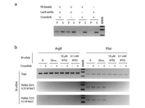
Stability of the LacI-LacO interaction. LacI-DNA complexes were prepared from E. coli K12 cells submitted or not to chemical crosslinking using formaldehyde, and the persistence of complexes was tested after two day-long washing. (a) Plac DNA fragment was immunoprecipitated by incubation of LacI antibody with an excess of cellular extracts (P, pellet; S, supernatant). Plac immunoprecipitation was no longer obtained in absence of protein A-coated beads (PA-beads), ruling out nonspecific adsorption to tube surfaces, and in absence of LacI antibody, ruling out nonspecific binding to the beads. (b) The persistence of LacI-Plac interaction was tested by high stringency washing of the DNA protein complexes attached to PA beads. A total of 4 tube transfers and 10 cycles of supernatant removal and replacement with fresh washing medium, were used for the final condition. Fig 2. PMID:?23454080
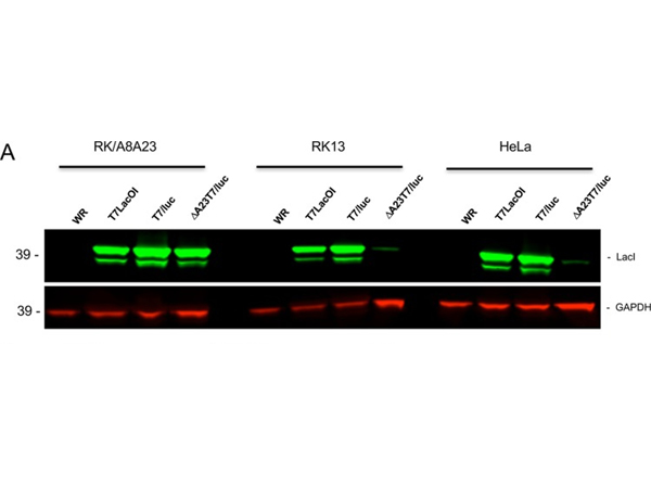
?(A) RK/A8A23, RK13, and HeLa cells were infected with VACV WR (WR), T7LacOI, or T7/luc replication-competent virus or with replication-defective ΔA23T7/luc virus at a multiplicity of 5?PFU/cell. After 24?h, the cells were lysed and LacI was detected by SDS polyacrylamide gel electrophoresis followed by immunoblotting and probing with specific antibody. Antibody to GAPDH was used as a loading control.?Fig 5. PMID:?28588133
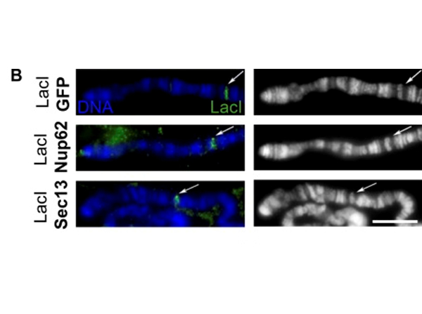
(B)?Widefield IF of squashed polytene chromosomes with Hoechst stain (labeled DNA and shown as blue or white/gray here and hereafter) and α-LacI (green). The right column shows Hoechst only in grayscale, whereas the left column shows the overlay of both channels. Arrows point to the?lacO?integration site at location 4D5 near the end of the X chromosome. LacI-fusion protein expression was driven with second chromosome Nubbin-Gal4. The scale bar is 10 μm. Fig 1. PMID:?31366666
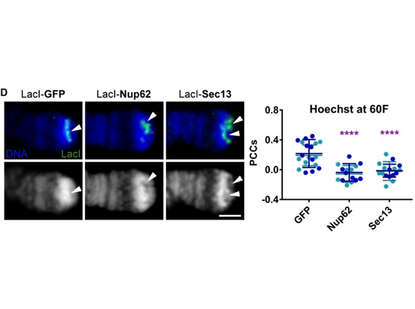
(D)?Confocal IF images of LacI-fusion proteins targeted to the subtelomeric?lacO?integration site on squashed polytene chromosomes at location 60F stained with Hoechst (blue or white) and α-LacI (green). The top row shows the overlay of both channels, and the bottom row shows Hoechst only. “Holes” (areas of highly reduced staining density) in Hoechst staining can be reproducibly observed under LacI-Nup binding. Expression was driven with Sgs3-Gal4. Arrowheads show observed decondensation or lack thereof under LacI. The scale bar is 2 μm. Quantification displays PCCs between blue and green signal under LacI. Data are from two biological replicates (colored), each from an independent experiment. GFP,?n?= 19; Nup62,?n?= 15; Sec13,?n?= 17. ****, P < 0.0001. Error bars represent SDs. Fig 1. PMID:?31366666
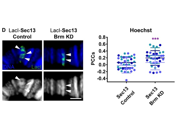
?(D)?Confocal IF images of LacI-Sec13 targeted to the?lacO?integration site on squashed polytene chromosomes at location 96C under control conditions (flies crossed to w1118 WT stock) or Brm KD conditions (flies crossed to Brm RNAi stock BL35211). Staining was with Hoechst (blue or white) and α-LacI (green). LacI-Sec13 protein expression and Brm RNAi were driven with Nubbin-Gal4. The top row shows the overlay of the two channels, whereas the bottom row shows DNA stain only in white/grayscale. Arrowheads indicate locations of observed decondensation or lack thereof under LacI signal. The scale bar is 2 μm. Quantification displays PCCs between red and blue signal under LacI. Data are from three biological replicates (colored) from two independent experiments. Sec13,?n?= 33. ***, P < 0.001. Error bars represent SDs. Fig 5. PMID:?31366666
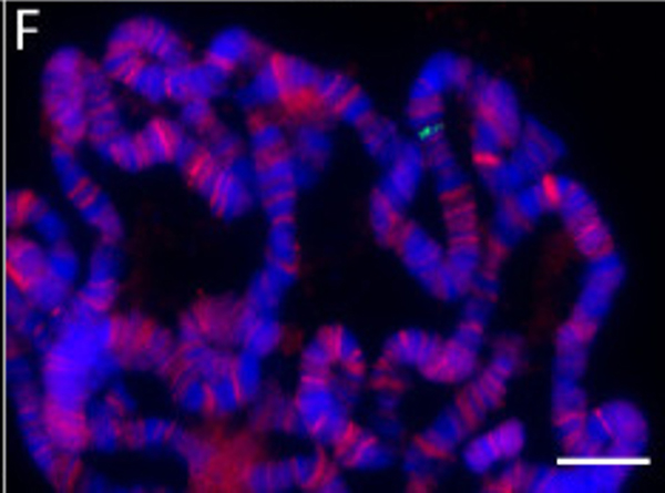
?F. Ideal squash and stain. Example includes?lacO?integration site at cytological site 96C, staining with antibodies against Nup98 (red, originally described in?Capelson?et al., 2010?) and LacI (green, Rockland 600-401-B04), recognizing LacI-Sec13 fusion protein bound to?lacO?96C, and Hoechst staining (blue). Scale bar = 10 μm. Fig 3. PMID:?33659343
|
|
|
|
Western Blot of Rabbit Anti-Lac I Antibody. Lane 1: Molecular Weight Marker (3μL). Lane 2: recombinant LAC-I (0.05μg). Primary Antibody: Anti-LAC-I at 1:1000 for 1hr at RT. Secondary Antibody: Goat anti-Rabbit IgG HRP (p/n 611-103-122) at 1:40,000 for 30mins at RT. Block: BlockOut Buffer (p/n MB-073) for 30mins at RT. Predicted/Observed MW: ~38kDa.
|
|
| 別品名 |
rabbit anti-LacI antibody, rabbit anti-Lac-I antibody, Rabbit anti-Lac I antibody, Lactose operon repressor, DNA binding transcriptional repressor antibody, Transcriptional repressor of the lac operon antibody
|
| 別包装 |
あり
|
| 交差種 |
Escherichia coli
Shigella
|
| 適用 |
Western Blot
Enzyme Linked Immunosorbent Assay
|
| 免疫動物 |
Rabbit
|
| 標識物 |
Unlabeled
|
| 精製度 |
Affinity Purified
|
| GENE ID |
8156885
|
| Accession No.(Gene/Protein) |
146576, P03023
|
| Gene Symbol |
LacI
|
| 参考文献 |
[[Cosmo Reference Collection]][Pub Med ID]22523743,23454080,28588133,31366666,33659343
|
| [注意事項] |
濃度はロットによって異なる可能性があります。メーカーDS及びCoAからご確認ください。
|
|
| メーカー |
品番 |
包装 |
|
RKL
|
600-401-B04
|
100 UG
|
※表示価格について
| 当社在庫 |
なし
|
| 納期目安 |
約10日
|
| 保存温度 |
-20℃
|
|


