|
※サムネイル画像をクリックすると拡大画像が表示されます。
Figure 1 Western Blot Validation of PD-L1 in HeLa Cells
Loading: 15 μg of lysates per lane.Antibodies: 4059 (A: 1 μg/mL, B: 2 μg/mL), 1 h incubation at RT in 5% NFDM/TBST.Secondary: Goat anti-rabbit IgG HRP conjugate at 1:10000 dilution.
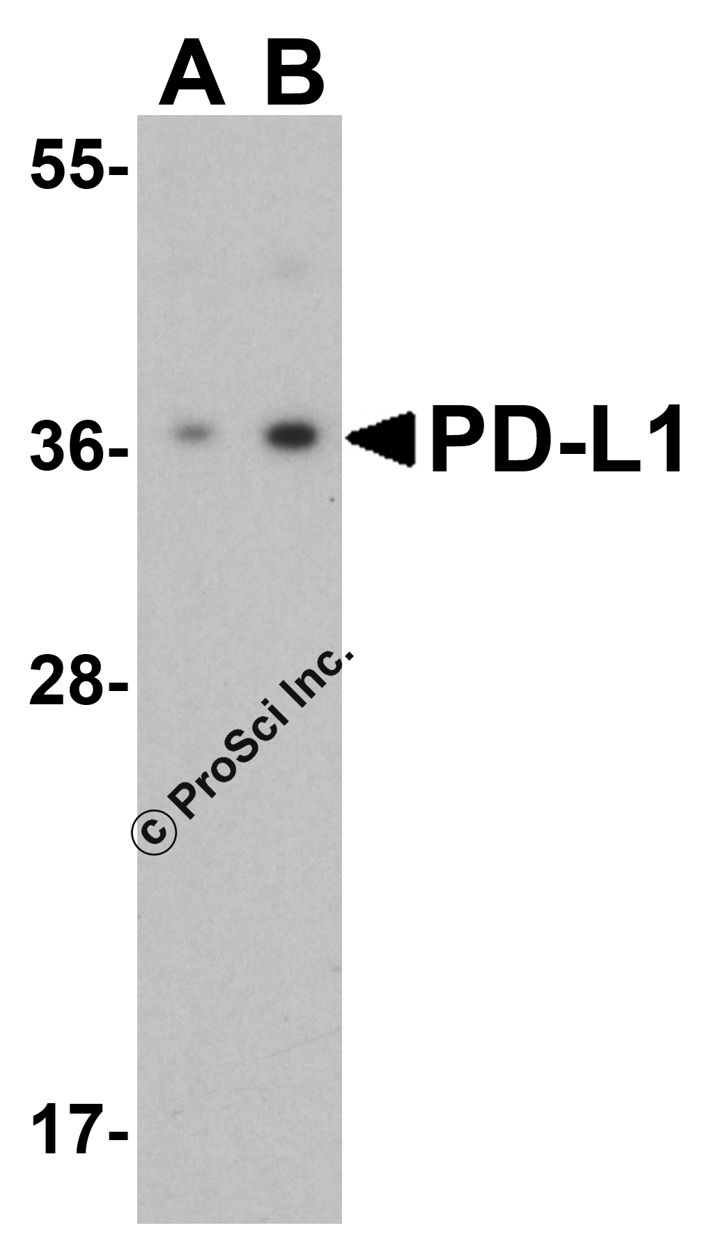
Figure 2 Independent Antibody Validation (IAV) via Protein Expression Profile in Human and Mouse cell lines
Loading: 15 μg of lysates per lane.Antibodies: 4059 (2 μg/mL), RF16035 (2 μg/mL), and beta-actin (1 μg/mL), 1 h incubation at RT in 5% NFDM/TBST.Secondary: Goat anti-rabbit and or anti-mouse IgG HRP conjugate at 1:10000 and 1:5000 dilution, respectively.
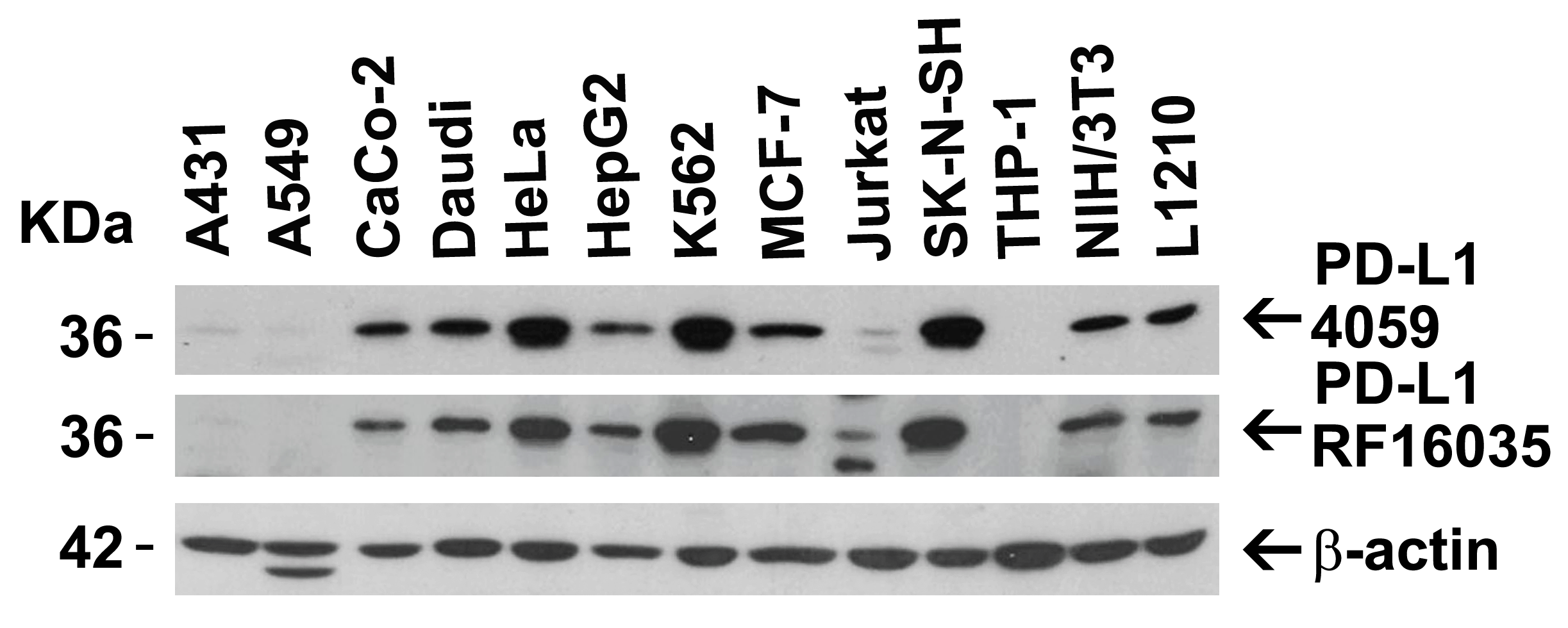
Figure 3 Validation with PD-L1 siRNA Knockdown in HeLa Cells
HeLa cells were transfected with control siRNAs (lane 1) or PD-L1 siRNAs (lane 2) Loading: 10 μg of HeLa whole cell lysates per lane.Antibodies: 4059 (2 μg/mL) and GAPDH (3783, 0.02 μg/mL), 1 h incubation at RT in 5% NFDM/TBST.Secondary: Goat anti-mouse IgG HRP conjugate at 1:5000 dilution.
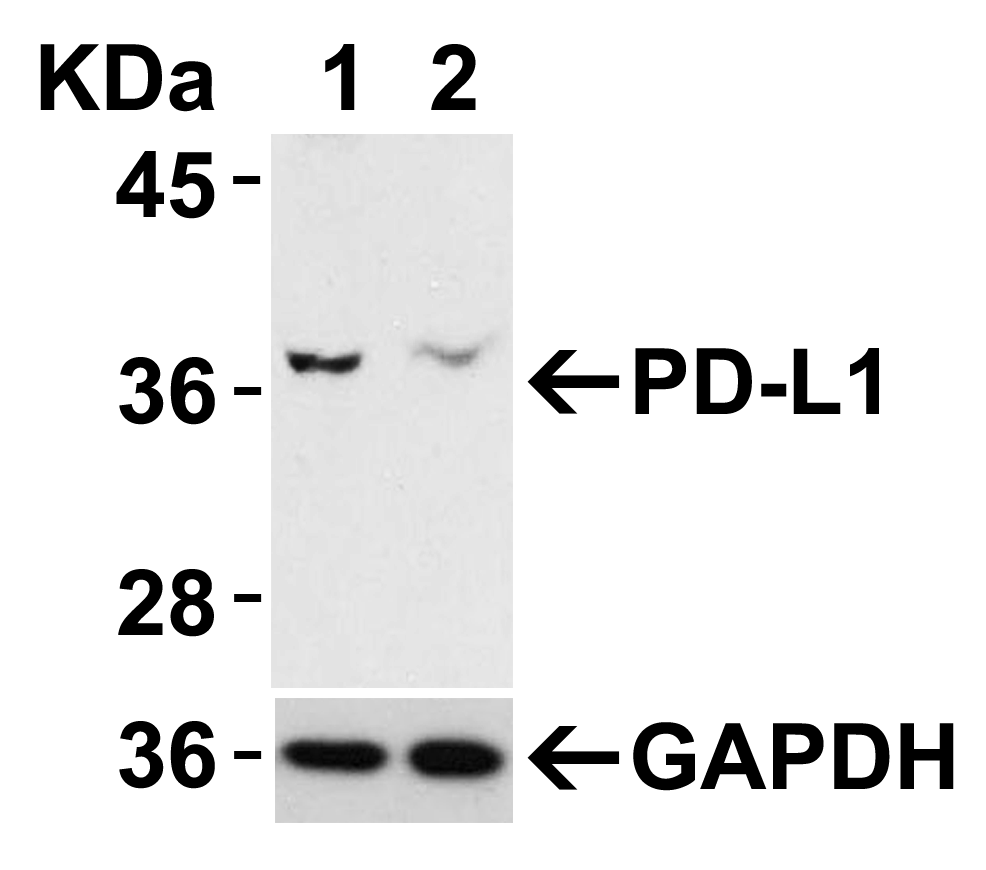
Figure 4 Validation with PD-L1 overexpression in 293 cells
Loading: Lysates/proteins at 15 μg per lane.Lane 1: non-transfected 293 cellsLane 2: PD-L1 overexpressed 293 cellsAntibodies: 4059 (1 μg/mL). 1 h incubation at RT in 5% NFDM/TBST.Secondary: Goat anti-rabbit IgG HRP conjugate at 1:10000 dilution.
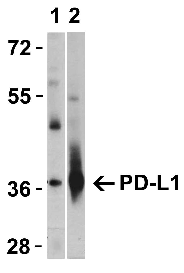
Figure 5 Immunohistochemistry Validation of PD-L1 in Human Tonsil Cells
Immunohistochemical analysis of paraffin-embedded human tonsil tissue using anti-PD-L1 antibody (4059) at 5 μg/ml. Tissue was fixed with formaldehyde and blocked with 10% serum for 1 h at RT; antigen retrieval was by heat mediation with a citrate buffer (pH6). Samples were incubated with primary antibody overnight at 4℃. A goat anti-rabbit IgG H&L (HRP) at 1/250 was used as secondary. Counter stained with Hematoxylin.
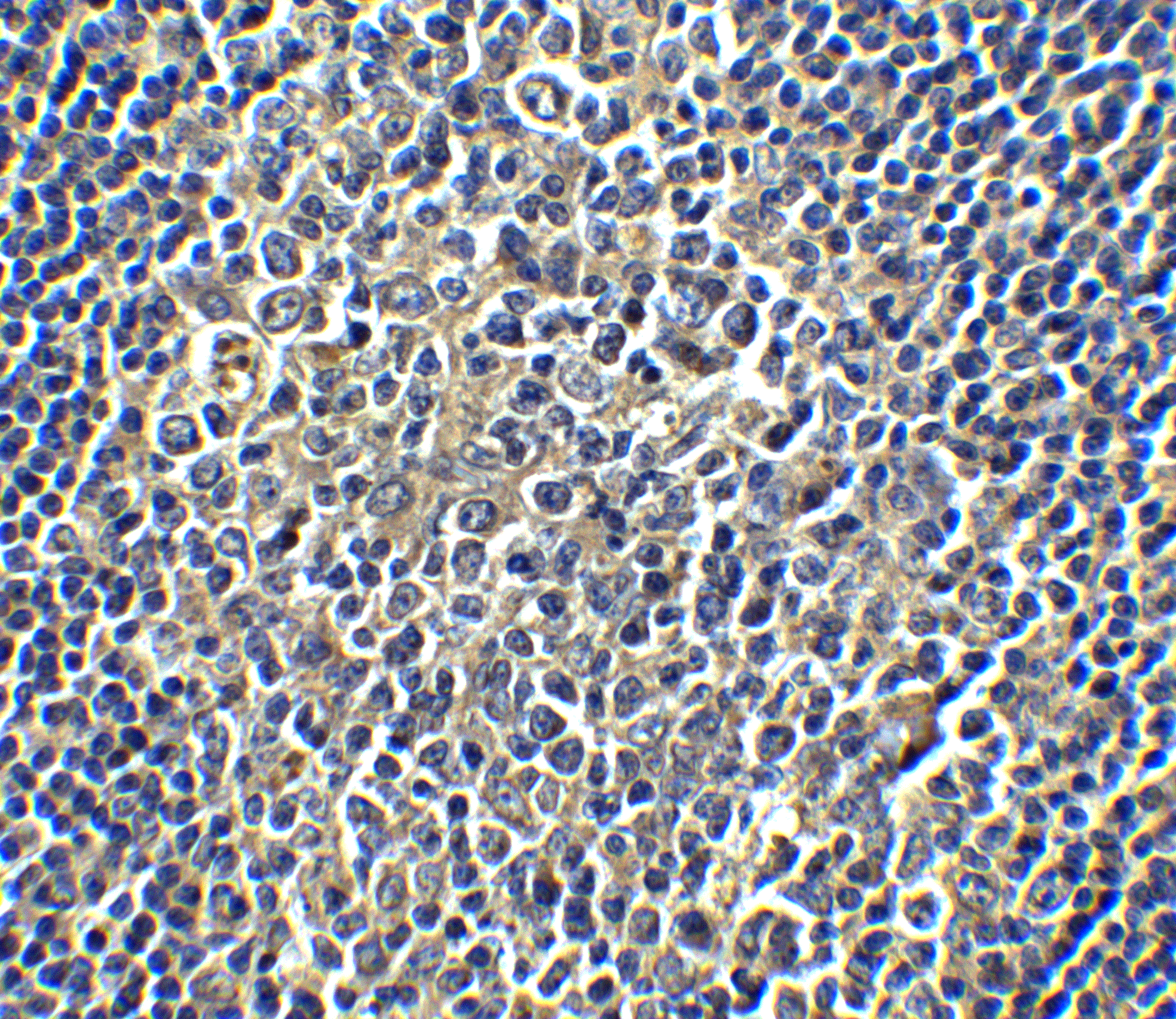
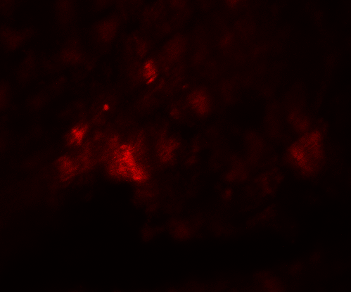
Figure 6 Immunofluorescence Validation of PD-L1 in Human Heart
Immunofluorescent analysis of 4% paraformaldehyde-fixed human heart tissue labeling PD-L1 with 4059 at 20 μg/mL, followed by goat anti-rabbit IgG secondary antibody at 1/500 dilution (red). Image showing both membrane and cytoplasmic staining on human heart tissue.
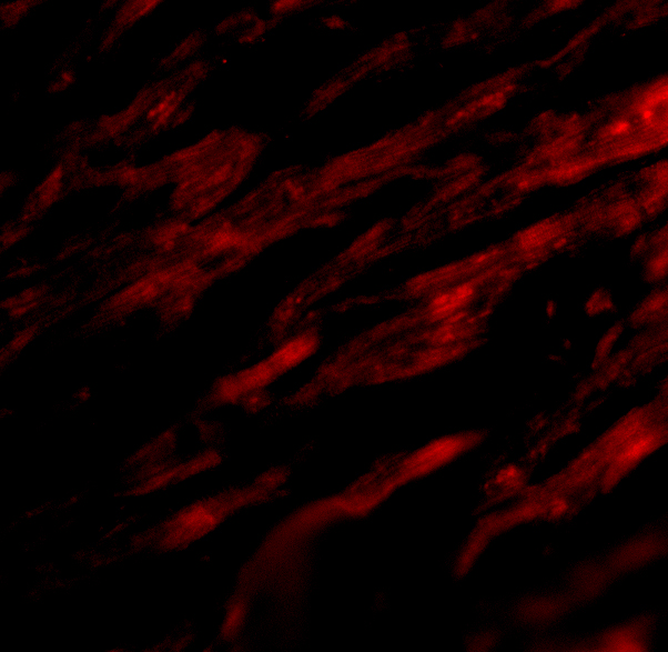
Figure 7 Flow Cytometry Validation of PD-L1
Overlay histogram showing A-20 cells stained with 4059 (red line, 1μg/1x106 cells). 1 h incubation at 4℃ in 2% FBS/PBS. Followed by secondary antibody 488 goat anti-rabbit IgG (H+L) at 1/500 dilution for 1 h 4℃. Isotype control antibody (Green line) was mouse IgG1 (1μg/1x106 cells) used under the same conditions. Acquisition of >10,000 events was performed.
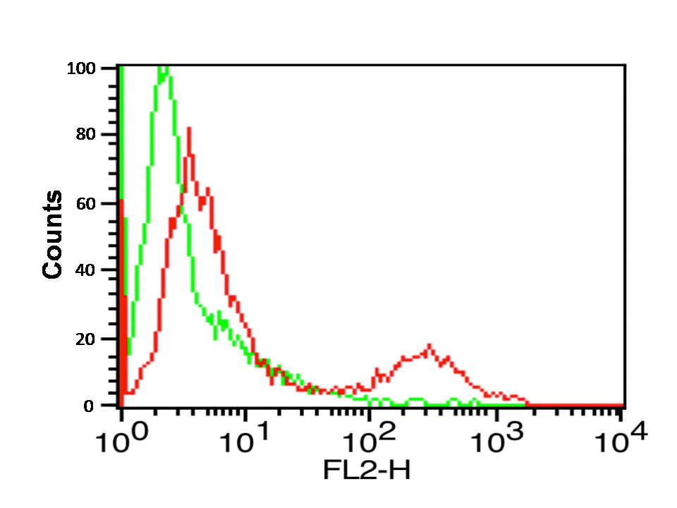
Figure 8 Immunohistochemistry Validation of PD-L1 in Rat Heart
Immunohistochemical analysis of paraffin-embedded rat heart tissue using anti-PD-L1 antibody (4059) at 5 μg/ml. Tissue was fixed with formaldehyde and blocked with 10% serum for 1 h at RT; antigen retrieval was by heat mediation with a citrate buffer (pH6). Samples were incubated with primary antibody overnight at 4℃. A goat anti-rabbit IgG H&L (HRP) at 1/250 was used as secondary. Counter stained with Hematoxylin.
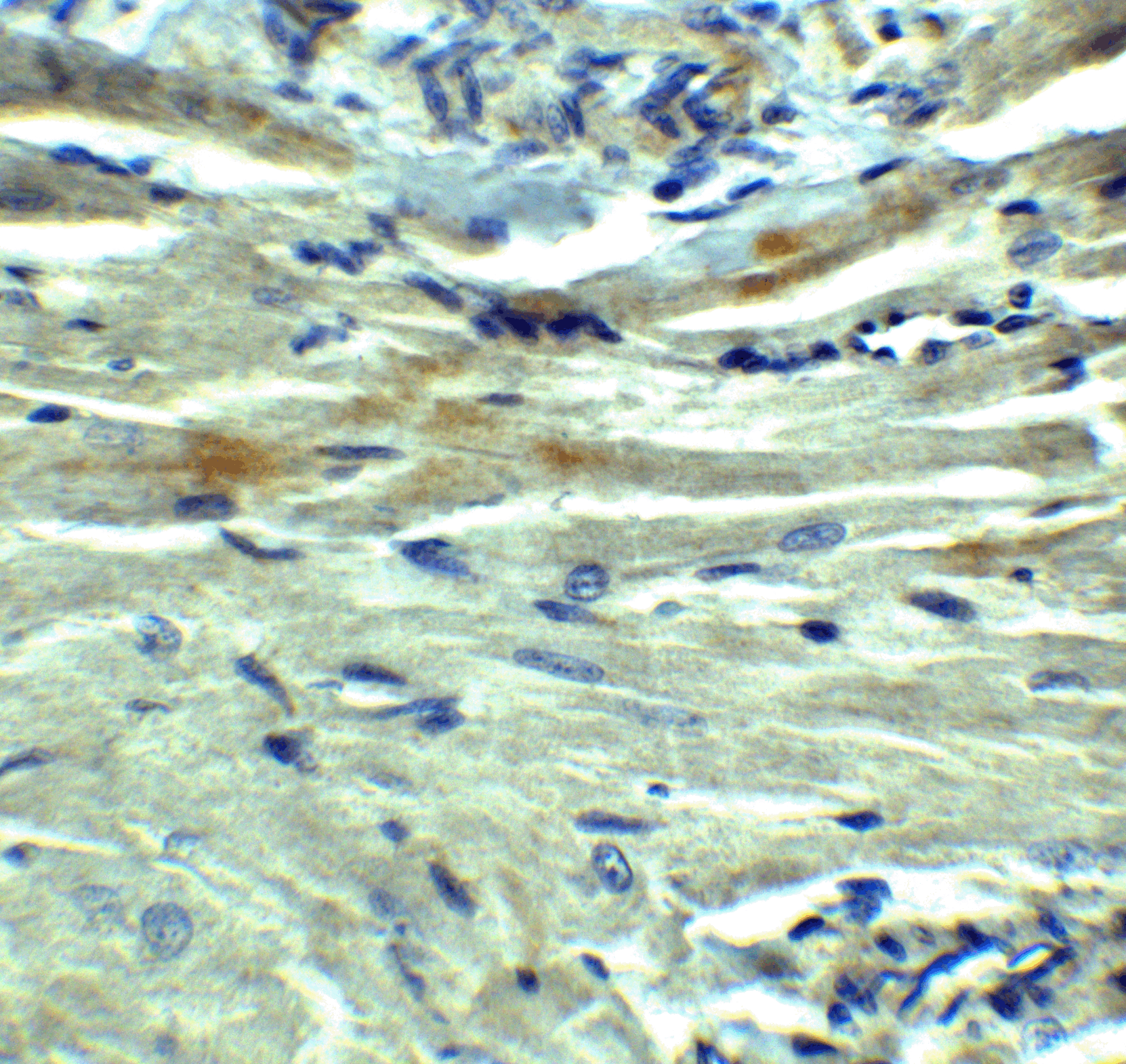
Figure 9 Immunohistochemistry Validation of PD-L1 in Human Heart
Immunohistochemical analysis of paraffin-embedded human heart tissue using anti-PD-L1 antibody (4059) at 2.5 μkg/ml. Tissue was fixed with formaldehyde and blocked with 10% serum for 1 h at RT; antigen retrieval was by heat mediation with a citrate buffer (pH6). Samples were incubated with primary antibody overnight at 4℃. A goat anti-rabbit IgG H&L (HRP) at 1/250 was used as secondary. Counter stained with Hematoxylin.
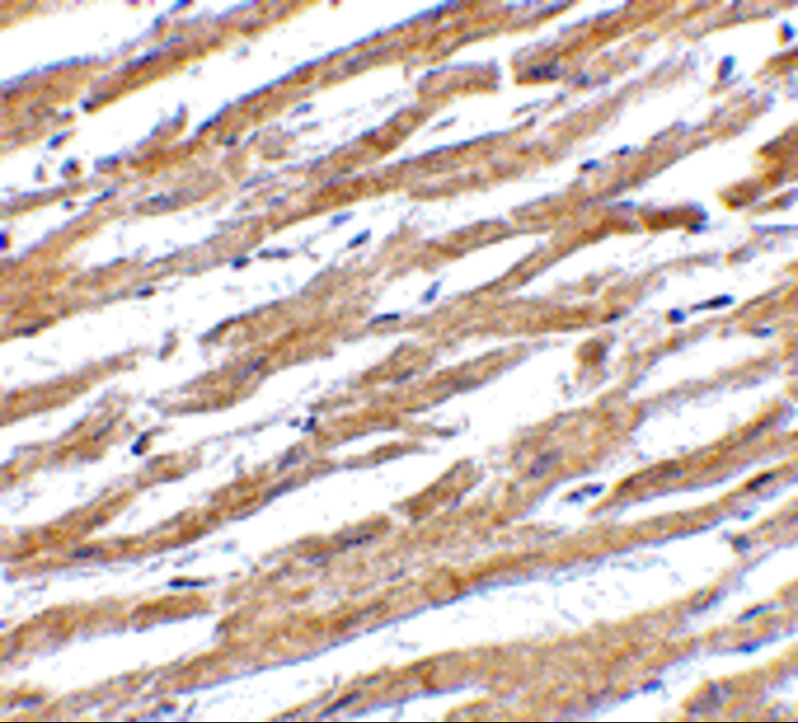
Figure 10 Immunofluorescence Validation of PD-L1 in Rat Heart
Immunofluorescence analysis of 4% paraformaldehyde-fixed rat heart tissue labeling PD-L1 with 4059 at 20 μg/ml, followed by goat anti-rabbit IgG secondary antibody at 1/250 dilution (red).
|
|
|
|
Figure 1 Western Blot Validation of PD-L1 in HeLa Cells
Loading: 15 μg of lysates per lane.Antibodies: 4059 (A: 1 μg/mL, B: 2 μg/mL), 1 h incubation at RT in 5% NFDM/TBST.Secondary: Goat anti-rabbit IgG HRP conjugate at 1:10000 dilution.
|
|
| 別品名 |
PD-L1 Antibody: B7-H, B7H1, PDL1, PD-L1, PDCD1L1, PDCD1LG1, Programmed cell death 1 ligand 1, B7 homolog 1
|
| 交差種 |
Human
Mouse
Rat
|
| 適用 |
Western Blot
IHC paraffin embedding section
Enzyme Linked Immunosorbent Assay
Immuno Fluorescence
Flow Cytometry
|
| 免疫動物 |
Rabbit
|
| 抗体クラス |
IgG
|
| 標識物 |
Unlabeled
|
| 精製度 |
Affinity Purified
|
| GENE ID |
29126
|
| Accession No.(Gene/Protein) |
NP_054862
|
| Gene Symbol |
CD274
|
| 推奨品 |
ポジティブコントロール 品番:1207 - Raji Cell Lysate
ポジティブコントロール 品番:1301 - Human Heart Tissue Lysate
|
| その他 |
[Protein GI Number]7661534
[Swiss-Prot No]Q9NZQ7
|
| 参考文献 |
Holling TM, Schooten E, and van Den Elsing PJ. Function and regulation of MHC class II molecules in T-lymphocytes: of mice and men. Hum. Immunol. 2004; 65:282-90.
Ishida Y, Agata Y, Shibahara K, et al. Induced expression of PD-1, a novel member of the immunoglobulin gene superfamily, upon programmed cell death. EMBO J. 1992; 11:3887-95.
LaGier J and Pober JS. Immune accessory functions of human endothelial cells are modulated by overexpression of B7-H1 (PDL1). Hum. Immunol. 2006; 67:568-78.
|
|
| メーカー |
品番 |
包装 |
|
PSC
|
4059
|
100 UG
[1 mg/mL]
|
※表示価格について
| 当社在庫 |
なし
|
| 納期目安 |
3週間程度
|
| 保存温度 |
-20℃
|
|
※当社では商品情報の適切な管理に努めておりますが、表示される法規制情報は最新でない可能性があります。
また法規制情報の表示が無いものは、必ずしも法規制に非該当であることを示すものではありません。
商品のお届け前に最新の製品法規制情報をお求めの際はこちらへお問い合わせください。
|
※当社取り扱いの試薬・機器製品および受託サービス・創薬支援サービス(納品物、解析データ等)は、研究用としてのみ販売しております。
人や動物の医療用・臨床診断用・食品用としては、使用しないように、十分ご注意ください。
法規制欄に体外診断用医薬品と記載のものは除きます。
|
|
※リンク先での文献等のダウンロードに際しましては、掲載元の規約遵守をお願いします。
|
|
※CAS Registry Numbers have not been verified by CAS and may be inaccurate.
|
