|
※サムネイル画像をクリックすると拡大画像が表示されます。
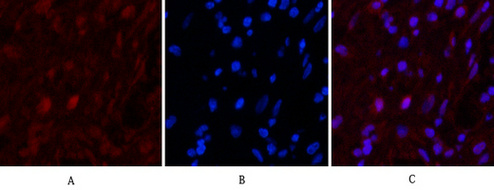
Immunofluorescence analysis of Human-appendix tissue. 1,Caspase 9 Monoclonal Antibody(3-20)(red) was diluted at 1:200(4°C,overnight). 2, Cy3 labled Secondary antibody was diluted at 1:300(room temperature, 50min).3, Picture B: DAPI(blue) 10min. Picture A:Target. Picture B: DAPI. Picture C: merge of A+B
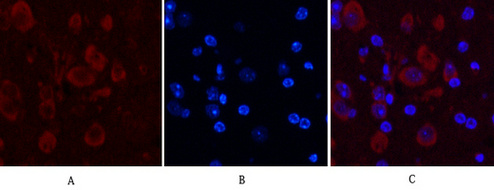
Immunofluorescence analysis of Mouse-brain tissue. 1,Caspase 9 Monoclonal Antibody(3-20)(red) was diluted at 1:200(4°C,overnight). 2, Cy3 labled Secondary antibody was diluted at 1:300(room temperature, 50min).3, Picture B: DAPI(blue) 10min. Picture A:Target. Picture B: DAPI. Picture C: merge of A+B
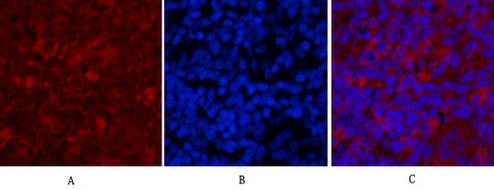
Immunofluorescence analysis of Rat-spleen tissue. 1,Caspase 9 Monoclonal Antibody(3-20)(red) was diluted at 1:200(4°C,overnight). 2, Cy3 labled Secondary antibody was diluted at 1:300(room temperature, 50min).3, Picture B: DAPI(blue) 10min. Picture A:Target. Picture B: DAPI. Picture C: merge of A+B
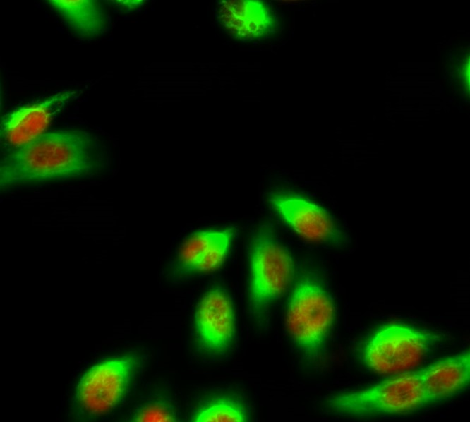
Immunofluorescence analysis of Hela cell. 1,eIF2α Polyclonal Antibody(red) was diluted at 1:200(4° overnight). Caspase 9 Monoclonal Antibody(3-20)(green) was diluted at 1:200(4° overnight). 2, Goat Anti Rabbit Alexa Fluor 594 Catalog:SA0837 was diluted at 1:1000(room temperature, 50min). Goat Anti Mouse Alexa Fluor 488 Catalog:SA0662 was diluted at 1:1000(room temperature, 50min).
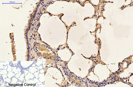
Immunohistochemical analysis of paraffin-embedded Human-lung tissue. 1,Caspase 9 Monoclonal Antibody(3-20) was diluted at 1:200(4°C,overnight). 2, Sodium citrate pH 6.0 was used for antibody retrieval(>98°C,20min). 3,Secondary antibody was diluted at 1:200(room tempeRature, 30min). Negative control was used by secondary antibody only.
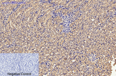
Immunohistochemical analysis of paraffin-embedded Mouse-kidney tissue. 1,Caspase 9 Monoclonal Antibody(3-20) was diluted at 1:200(4°C,overnight). 2, Sodium citrate pH 6.0 was used for antibody retrieval(>98°C,20min). 3,Secondary antibody was diluted at 1:200(room tempeRature, 30min). Negative control was used by secondary antibody only.
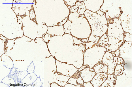
Immunohistochemical analysis of paraffin-embedded Rat-lung tissue. 1,Caspase 9 Monoclonal Antibody(3-20) was diluted at 1:200(4°C,overnight). 2, Sodium citrate pH 6.0 was used for antibody retrieval(>98°C,20min). 3,Secondary antibody was diluted at 1:200(room tempeRature, 30min). Negative control was used by secondary antibody only.
1) Input: Hela Cell Lysate 2) IP product: IP dilute 1:200
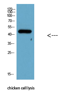
Western Blot analysis of chicken cell lysis using Antibody diluted at 1:1000
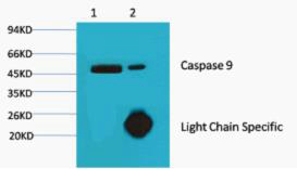
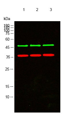
Western blot analysis of lysates from 1) Hela, 2) Jurkat, 3)3T3 cells, (Green) primary antibody was diluted at 1:1000, 4°over night, secondary antibody(cat:SA0438)was diluted at 1:10000, 37° 1hour. (Red) GAPDH Polyclonal Antibody (cat:YM3215) antibody was diluted at 1:5000 as loading control, 4° over night,secondary antibody(cat:SA0437)was diluted at 1:10000, 37° 1hour.
|
|
|
|
Immunofluorescence analysis of Human-appendix tissue. 1,Caspase 9 Monoclonal Antibody(3-20)(red) was diluted at 1:200(4°C,overnight). 2, Cy3 labled Secondary antibody was diluted at 1:300(room temperature, 50min).3, Picture B: DAPI(blue) 10min. Picture A:Target. Picture B: DAPI. Picture C: merge of A+B
|
|
| 別品名 |
CASP9; MCH6; Caspase-9; CASP-9; Apoptotic protease Mch-6; Apoptotic protease-activating factor 3; APAF-3; ICE-like apoptotic protease 6; ICE-LAP6
|
| 交差種 |
Human
Mouse
Rat
Chicken
|
| 適用 |
Western Blot
IHC paraffin embedding section
Immuno Fluorescence
Immunocytochemistry (cell)
Immunoprecipitation
|
| 免疫動物 |
Mouse
|
| Accession No.(Gene/Protein) |
P55211
|
|
| メーカー |
品番 |
包装 |
|
ASY
|
N1342
|
100 UL
|
※表示価格について
| 当社在庫 |
なし
|
| 納期目安 |
2週間程度
|
| 保存温度 |
-20℃
|
|
※当社では商品情報の適切な管理に努めておりますが、表示される法規制情報は最新でない可能性があります。
また法規制情報の表示が無いものは、必ずしも法規制に非該当であることを示すものではありません。
商品のお届け前に最新の製品法規制情報をお求めの際はこちらへお問い合わせください。
|
※当社取り扱いの試薬・機器製品および受託サービス・創薬支援サービス(納品物、解析データ等)は、研究用としてのみ販売しております。
人や動物の医療用・臨床診断用・食品用としては、使用しないように、十分ご注意ください。
法規制欄に体外診断用医薬品と記載のものは除きます。
|
|
※リンク先での文献等のダウンロードに際しましては、掲載元の規約遵守をお願いします。
|
|
※CAS Registry Numbers have not been verified by CAS and may be inaccurate.
|
