|
※サムネイル画像をクリックすると拡大画像が表示されます。
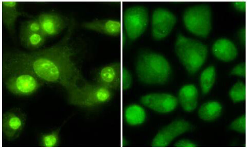
Immunofluorescence microscopy anti -THRB1 (Thyroid hormone receptor Beta 1) antibody 600-401-A96. Tissue: Mouse Dendritic cells. Primary antibody: Anti THRB1 1:100 1 hr PBS 3% BSA (left) Normal rabbit IgG isotype control (right). Secondary Ab: 488 dye conjugate 1:1000 1 hr. Mounting: Fluoromount-G (Southern Biotechnology Associates, Birmingham, AL) for examination. This image appeared originally in Mascanfroni, Ivan D ; del Mar Montesinos M; Alamino Vanina A. ; Susperreguy S, Nicola JP, Ilarregui JM, Masini-Repiso AM, Rabinovich GA, Pellizas CG (2010) Nuclear Factor (NF)-kappa B-dependent Thyroid Hormone Receptor beta(1) Expression Controls Dendritic Cell Function via Akt Signaling. Journal of Biological Chemistry 285 (13), 9569-9582. DOI: 10.1074/jbc.M109.071241 Published: MAR 26 2010. Copyright c 2010, by the American Society for Biochemistry and Molecular Biology.
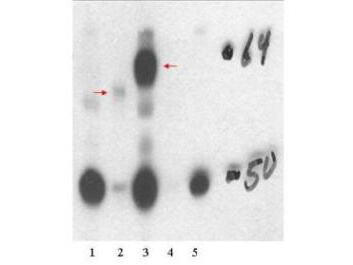
Western blot using Rockland's affinity purified anti-THRB1 antibody shows detection of in vitro translated THRB1 (lane 2) and human thyroid stable cell line expressing THTRb1 (lane 3). No staining is evident in non transfected human thyroid cells (lane 1), over expressed THRA (lane 4) or HeLa stable cell line expressing THRA1 (lane 5). The band at ~62 kDa, indicated by the arrowheads, corresponds to THRB. Personal communication, H. Ying, NCI, Bethesda, MD
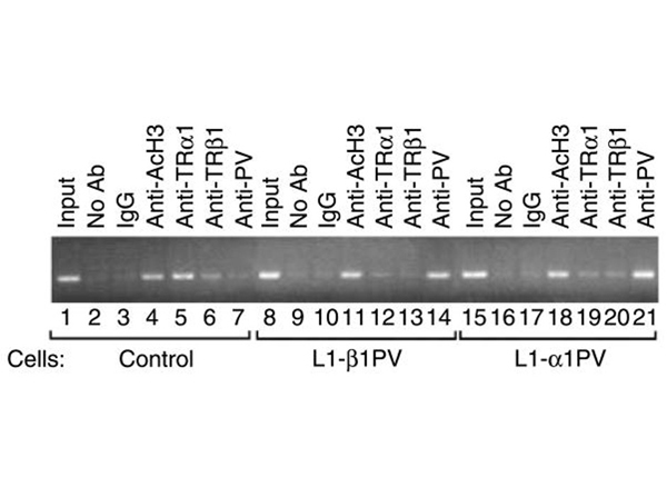
Differential recruitment of TRβ1PV and TRα1PV to the promoter of the?C/ebpα?gene as determined by ChIP analysis. ChIP assays were performed using control 3T3-L1 (lanes 1?7), L1-β1PV (lanes 8?14), and L1-α1PV (lanes 15?21) cells after T3-induced adipogenesis on day 9. Antibodies used in the chromatin immunoprecipitation were anti-Ac-H3 antibody as a positive control (lanes 4, 11, and 18), anti-TRα1 (p/n 600-401-A38) antibody (lanes 5, 12, and 19), anti-TRβ1 (p/n 600-401-A96) antibody (lanes 6, 13, and 20), and anti-PV antibody (lanes 7, 14, and 21). The negative controls were no antibody (lanes 2, 9, and 16) as well as an irrelevant IgG (lanes 3, 10, and 17). The chromatin immunoprecipitated and recovered DNA was used as a template for PCR amplification of the receptor-binding region in the promoter of the?C/ebpα?gene. Two percent of the chromatin solution (20 μl) was used for input DNA as a control. Three separate experiments were performed, and the representative results are shown. Fig 6. PMID:?20080985
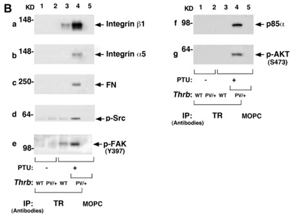
B, Coimmunoprecipitation assays were carried out as described in?Materials and Methods. PV physically interacted with integrin-β1 (panel a), integrin-α5 (panel b), FN (panel c), p-Src (panel d), p-FAK (panel e), p85α (panel f), and p-AKT (panel g). An increased association of PV with the indicated regulators was observed in the thyroid of the?ThrbPV/+-PTU mice (lane 4) as compared with the WT-PTU mice (lane 3). No apparent association of PV with the indicated key regulators was apparent in the thyroid of the untreated?ThrbPV/+(lane 2) mice. Lane 5 shows the negative control when an irrelevant antibody, mouse monoclonal IgG MOPC-141, was used in the immunoprecipitation. Fig 7. PMID:?22919057
|
|
|
|
Immunofluorescence microscopy anti -THRB1 (Thyroid hormone receptor Beta 1) antibody 600-401-A96. Tissue: Mouse Dendritic cells. Primary antibody: Anti THRB1 1:100 1 hr PBS 3% BSA (left) Normal rabbit IgG isotype control (right). Secondary Ab: 488 dye conjugate 1:1000 1 hr. Mounting: Fluoromount-G (Southern Biotechnology Associates, Birmingham, AL) for examination. This image appeared originally in Mascanfroni, Ivan D ; del Mar Montesinos M; Alamino Vanina A. ; Susperreguy S, Nicola JP, Ilarregui JM, Masini-Repiso AM, Rabinovich GA, Pellizas CG (2010) Nuclear Factor (NF)-kappa B-dependent Thyroid Hormone Receptor beta(1) Expression Controls Dendritic Cell Function via Akt Signaling. Journal of Biological Chemistry 285 (13), 9569-9582. DOI: 10.1074/jbc.M109.071241 Published: MAR 26 2010. Copyright c 2010, by the American Society for Biochemistry and Molecular Biology.
|
|
| 別品名 |
Rabbit anti-THRB antibody, rabbit anti-Thyroid hormone receptor beta 1 antibody, Anti-THRB1 antibody, c-erbA-beta, THR1 antibody, c-erbA-2, Nuclear receptor subfamily 1 group A member 2, ERBA2, NR1A2
|
| 別包装 |
あり
|
| 交差種 |
Human
Mouse
|
| 適用 |
Western Blot
Enzyme Linked Immunosorbent Assay
Immuno Fluorescence
|
| 免疫動物 |
Rabbit
|
| 抗原部位 |
N-terminus
|
| 標識物 |
Unlabeled
|
| 精製度 |
Affinity Purified
|
| GENE ID |
7068
|
| Accession No.(Gene/Protein) |
40806162, P10828
|
| Gene Symbol |
THRB
|
| 参考文献 |
[[Cosmo Reference Collection]][Pub Med ID]20080985,22919057,23840792,27254276
|
| [注意事項] |
濃度はロットによって異なる可能性があります。メーカーDS及びCoAからご確認ください。
|
|
| メーカー |
品番 |
包装 |
|
RKL
|
600-401-A96S
|
25 UL
|
※表示価格について
| 当社在庫 |
なし
|
| 納期目安 |
約10日
|
| 保存温度 |
-20℃
|
|


