|
※サムネイル画像をクリックすると拡大画像が表示されます。
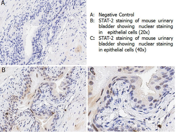
Immunohistochemistry with anti-STAT2 pY690 antibody showing nuclear positivity in epithelial cells of mouse urinary bladder tissue at 20x and 40x (B & C). Staining was performed on Leica Bond system using the standard protocol. Formalin fixed/paraffin embedded tissue sections were subjected to antigen retrieval and then incubated with rabbit anti-STAT2 pY690 antibody 600-401-A93 at 1:100 dilution for 60 minutes. Biotinylated Anti-rabbit secondary antibody was used at 1:200 dilution to detect primary antibody. The reaction was developed using streptavidin-HRP conjugated compact polymer system and visualized with chromogen substrate, 3’3-diamino-benzidine substrate (DAB). The sections were then counterstained with hematoxylin to detect cell nuclei.
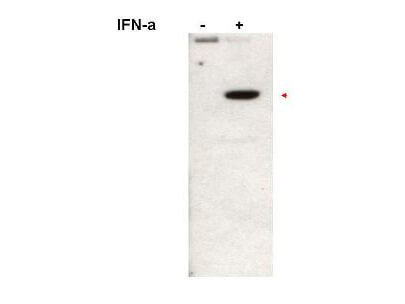
Western blot using Rockland Immunochemicals affinity purified anti-Stat2pY690 antibody shows detection of Stat2pY690 protein (arrowhead). Loaded Jurkat cells treated without (left lane) or with (right lane) 1000U/mL of IFN-a for 15 min at 37°C. Primary antibody was used at 1:1,000.? Personal Communication, A.Gamero, NCI, Bethesda, MD.
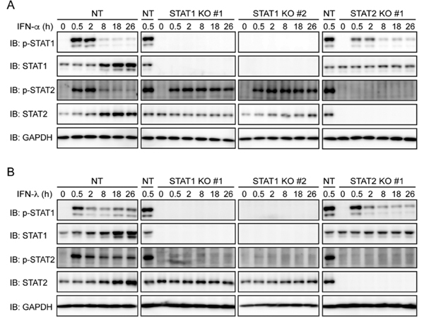
(A,B) NT sgRNA-expressing Huh-7.5 cells, STAT1 knockout (KO) cells (clones #1 and #2), and STAT2 KO cells (clone #1) were treated with IFN-α (1,000?U/ml) (A) or IFN-λ (1,000?U/ml) (B) for the indicated times. Phosphorylation of STAT1 and STAT2 was evaluated by immunoblotting (IB). Fig 7. PMID:?27929099
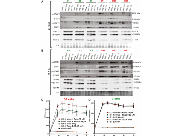
VSV and antiviral protein expression(A and B) C and GR cells were either mock treated or infected with VSV-ΔM51 at MOIs of 1.0, 0.1, and 0.01. Protein sample were analyzed at 24?h p.i. (A) and 48?h p.i. (B) by western blotting for expression of phospho-STAT1 (p-STAT1), phospho-STAT2 (p-STAT2), STAT1, STAT2, and VSV proteins (G, N/P, and M). Cell line and treatment conditions are indicated above blots. Equal protein loading is indicated by Coomassie blue. (C and D) The effect of ruxolitinib on VSV-ΔM51 replication kinetics based on GFP fluorescence. C and GR cells were either mock treated, treated only with ruxolitinib, treated only with VSV-ΔM51, or treated with both VSV-ΔM51 and ruxolitinib. Cells were infected at MOI of 0.01. Ruxolitinib was added to cells after 1?h VSV incubation period. GFP fluorescence was measured from 1 to 80?h p.i. The data points and error bars shown represent the means and SEM of the means, respectively. Control and GR cell lines 1?3 are combined for each treatment. Fig 5. PMID:?34977342
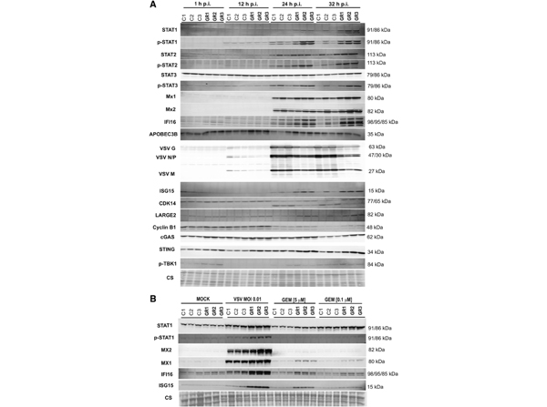
Time course expression of antiviral proteins in C and GR cells after VSV infection(A) C and GR cells were infected with VSV-ΔM51 at MOI 0.01. Protein samples were collected at 1, 12, 24, and 32?h p.i. and analyzed by western blotting. Cell lines and time points are indicted above the blots. Equal protein loading is indicated by Coomassie blue. (B) C and GR cells were either mock treated, infected with VSV-ΔM51, or treated with 5?μM gemcitabine or 0.1?μM gemcitabine. Protein samples were collected after 32?h and analyzed by western blotting for expression of ISGs. Cell lines and time points are indicted above the blots. Equal protein loading is indicated by Coomassie blue. Fig 7. PMID:?34977342
|
|
|
|
Immunohistochemistry with anti-STAT2 pY690 antibody showing nuclear positivity in epithelial cells of mouse urinary bladder tissue at 20x and 40x (B & C). Staining was performed on Leica Bond system using the standard protocol. Formalin fixed/paraffin embedded tissue sections were subjected to antigen retrieval and then incubated with rabbit anti-STAT2 pY690 antibody 600-401-A93 at 1:100 dilution for 60 minutes. Biotinylated Anti-rabbit secondary antibody was used at 1:200 dilution to detect primary antibody. The reaction was developed using streptavidin-HRP conjugated compact polymer system and visualized with chromogen substrate, 3’3-diamino-benzidine substrate (DAB). The sections were then counterstained with hematoxylin to detect cell nuclei.
|
|
| 別品名 |
rabbit anti-STAT2 pY690 antibody, phosphorylated STAT2 antibody, STAT-2, STAT 2, Signal transducer and activator of transcription 2, p113
|
| 別包装 |
あり
|
| 交差種 |
Human
Mouse
|
| 適用 |
Western Blot
Enzyme Linked Immunosorbent Assay
Immunohistochemistry
|
| 免疫動物 |
Rabbit
|
| 標識物 |
Unlabeled
|
| 精製度 |
Affinity Purified
|
| 翻訳後修飾 |
リン酸化
|
| GENE ID |
6773
|
| Accession No.(Gene/Protein) |
NP_005410.1, P52630
|
| Gene Symbol |
STAT2
|
| 参考文献 |
[[Cosmo Reference Collection]][Pub Med ID]27929099,34977342
|
| [注意事項] |
濃度はロットによって異なる可能性があります。メーカーDS及びCoAからご確認ください。
|
|
| メーカー |
品番 |
包装 |
|
RKL
|
600-401-A93S
|
25 UL
|
※表示価格について
| 当社在庫 |
なし
|
| 納期目安 |
約10日
|
| 保存温度 |
-20℃
|
|
※当社では商品情報の適切な管理に努めておりますが、表示される法規制情報は最新でない可能性があります。
また法規制情報の表示が無いものは、必ずしも法規制に非該当であることを示すものではありません。
商品のお届け前に最新の製品法規制情報をお求めの際はこちらへお問い合わせください。
|
※当社取り扱いの試薬・機器製品および受託サービス・創薬支援サービス(納品物、解析データ等)は、研究用としてのみ販売しております。
人や動物の医療用・臨床診断用・食品用としては、使用しないように、十分ご注意ください。
法規制欄に体外診断用医薬品と記載のものは除きます。
|
|
※リンク先での文献等のダウンロードに際しましては、掲載元の規約遵守をお願いします。
|
|
※CAS Registry Numbers have not been verified by CAS and may be inaccurate.
|


