|
※サムネイル画像をクリックすると拡大画像が表示されます。
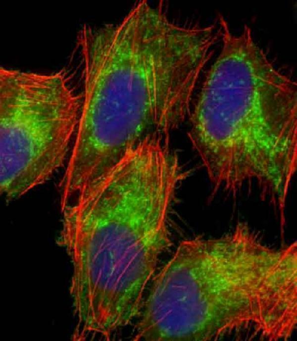
Immunofluorescent analysis of 4% paraformaldehyde-fixed, 0.1% Triton X-100 permeabilized U-2 OS (human osteosarcoma cell line) cells labeling TOMM40 with M03166 at 1/25 dilution, followed by DylightR 488-conjugated goat anti-rabbit IgG secondary antibody at 1/200 dilution (green). Immunofluorescence image showing mitochondrion staining on U-2 OS cell line. Cytoplasmic actin is detected with DylightR 554 Phalloidin at 1/100 dilution (red).The nuclear counter stain is DAPI (blue).
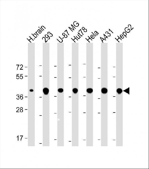
All lanes : Anti-TOMM40 Antibody (N-Term) at 1:2000 dilution
Lane 1: Human brain lysate
Lane 2: 293 whole cell lysate
Lane 3: U-87 MG whole cell lysate
Lane 4: Hut78 whole cell lysate
Lane 5: Hela whole cell lysate
Lane 6: A431 whole cell lysate
Lane 7: HepG2 whole cell lysate
Lysates/proteins at 20 μg per lane.
Secondary
Goat Anti-Rabbit IgG, (H+L), Peroxidase conjugated at 1/10000 dilution.
Predicted band size : 38 kDa
Blocking/Dilution buffer: 5% NFDM/TBST.
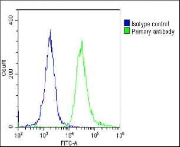
Overlay histogram showing HeLa cells stained with M03166 (green line). The cells were fixed with 2% paraformaldehyde (10 min) and then permeabilized with 90% methanol for 10 min. The cells were then icubated in 2% bovine serum albumin to block non-specific protein-protein interactions followed by the antibody (M03166, 1:25 dilution) for 60 min at 37oC. The secondary antibody used was Goat-Anti-Rabbit IgG, DyLightR 488 Conjugated Highly Cross-Adsorbed at 1/200 dilution for 40 min at 37oC. Isotype control antibody (blue line) was rabbit IgG1 (1g/1x10^6 cells) used under the same conditions. Acquisition of >10, 000 events was performed.
|
|
|
|
Immunofluorescent analysis of 4% paraformaldehyde-fixed, 0.1% Triton X-100 permeabilized U-2 OS (human osteosarcoma cell line) cells labeling TOMM40 with M03166 at 1/25 dilution, followed by DylightR 488-conjugated goat anti-rabbit IgG secondary antibody at 1/200 dilution (green). Immunofluorescence image showing mitochondrion staining on U-2 OS cell line. Cytoplasmic actin is detected with DylightR 554 Phalloidin at 1/100 dilution (red).The nuclear counter stain is DAPI (blue).
|
|
| 別品名 |
Mitochondrial import receptor subunit TOM40 homolog, Protein Haymaker, Translocase of outer membrane 40 kDa subunit homolog, p38.5, TOMM40, C19orf1, PEREC1, TOM40
|
| 種由来 |
Human
|
| 交差種 |
Human
|
| 適用 |
Western Blot
Immuno Fluorescence
Flow Cytometry
|
| 免疫動物 |
Rabbit
|
| 抗体クラス |
IgG
|
| 抗原部位 |
a.a.22-56
|
| 精製度 |
Ig fraction - Protein A
|
| Accession No.(Gene/Protein) |
O96008
|
| Gene Symbol |
TOMM40
|
| 分子量 |
37893 Da
|
| 概要 |
Boster Bio Anti-TOMM40 Antibody (N-Term) (Catalog # M03166). Tested in WB, Flow Cytometry, IF application(s). This antibody reacts with Human.
|
|
| メーカー |
品番 |
包装 |
|
BBT
|
M03166
|
50 UL
|
※表示価格について
| 当社在庫 |
なし
|
| 納期目安 |
1週間程度
|
| 保存温度 |
-20℃
|
|
※当社では商品情報の適切な管理に努めておりますが、表示される法規制情報は最新でない可能性があります。
また法規制情報の表示が無いものは、必ずしも法規制に非該当であることを示すものではありません。
商品のお届け前に最新の製品法規制情報をお求めの際はこちらへお問い合わせください。
|
※当社取り扱いの試薬・機器製品および受託サービス・創薬支援サービス(納品物、解析データ等)は、研究用としてのみ販売しております。
人や動物の医療用・臨床診断用・食品用としては、使用しないように、十分ご注意ください。
法規制欄に体外診断用医薬品と記載のものは除きます。
|
|
※リンク先での文献等のダウンロードに際しましては、掲載元の規約遵守をお願いします。
|
|
※CAS Registry Numbers have not been verified by CAS and may be inaccurate.
|
