
| 別品名 |
Interleukin-1 receptor-associated kinase 3, IRAK-3, IL-1 receptor-associated kinase M, IRAK-M, IRAK3 {ECO:0000312|EMBL:AAH578001}
|
| 抗原部位 |
a.a.45-77
|
| 種由来 |
Human
|
| 精製度 |
Ig fraction - Protein A
|
| 適用 |
IHC paraffin embedding section
Western Blot
|
| 免疫動物 |
Rabbit
|
| 抗体クラス |
IgG
|
| クローン |
RB2344
|
| 交差種 |
Human
Mouse
|
| GENE ID |
11213
|
| Accession No.(Gene/Protein) |
Q9Y616
|
| Gene Symbol |
IRAK3
|
| 分子量 |
67767
|
| 参考文献 |
Rosati, O., et al., Biochem. Biophys. Res. Commun. 293(5):1472-1477 (2002).Wesche, H., et al., J. Biol. Chem. 274(27):19403-19410 (1999).
|
|
※サムネイル画像をクリックすると拡大画像が表示されます。
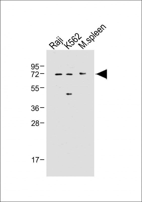
All lanes : Anti-IRAK3 Antibody (N-term) at 1:500 dilutionLane 1: Raji whole cell lysateLane 2: K562 whole cell lysateLane 3: Mouse spleen tissue lysateLysates/proteins at 20 μg per lane. SecondaryGoat Anti-Rabbit IgG, (H+L), Peroxidase conjugated at 1/10000 dilution. Predicted band size : 68 kDaBlocking/Dilution buffer: 5% NFDM/TBST.
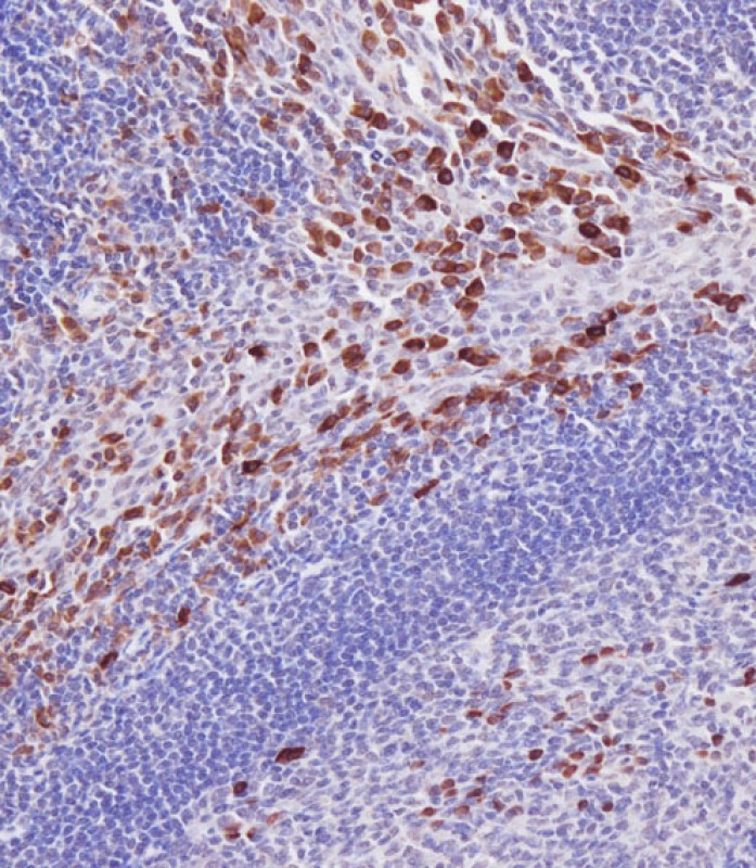
Immunohistochemical analysis of AP7804A on paraffin-embedded Human tonsil tissue. Tissue was fixed with formaldehyde at room temperature. Heat induced epitope retrieval was performed by EDTA buffer (pH9. 0). Samples were incubated with primary antibody(1:100) for 1 hour at room temperature. Undiluted CRF Anti-Polyvalent HRP Polymer antibody was used as the secondary antibody.
|
|
|
|
All lanes : Anti-IRAK3 Antibody (N-term) at 1:500 dilutionLane 1: Raji whole cell lysateLane 2: K562 whole cell lysateLane 3: Mouse spleen tissue lysateLysates/proteins at 20 μg per lane. SecondaryGoat Anti-Rabbit IgG, (H+L), Peroxidase conjugated at 1/10000 dilution. Predicted band size : 68 kDaBlocking/Dilution buffer: 5% NFDM/TBST.
|
|
|
| メーカー |
品番 |
包装 |
|
ABH
|
AP7804A-EV
|
80 UL
|
※表示価格について
|

