
| 別品名 |
Syndecan-1, SYND1, CD138, SDC1, SDC
|
| 種由来 |
Human
|
| 精製度 |
Ig fraction - Protein G
|
| 適用 |
Western Blot
Immuno Fluorescence
Flow Cytometry
|
| 免疫動物 |
Mouse
|
| 抗体クラス |
IgG1
|
| クローン |
480CT5.4.3
|
| 交差種 |
Human
|
| GENE ID |
6382
|
| Accession No.(Gene/Protein) |
P18827
|
| Gene Symbol |
SDC1
|
| 分子量 |
32462
|
|
※サムネイル画像をクリックすると拡大画像が表示されます。
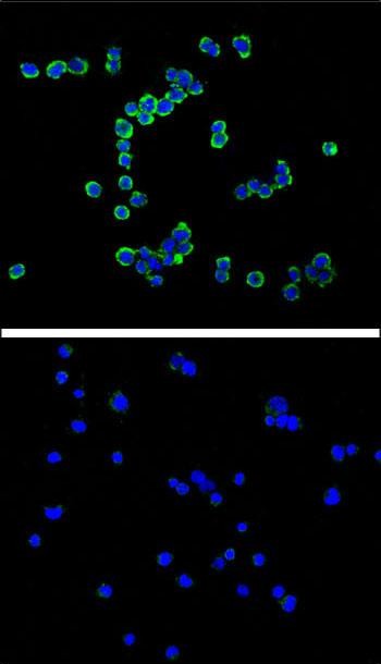
Confocal immunofluorescent analysis of CD138 antibody (Cat#AM2157a) with RPMI8266 cell (above) compared with Jurkat as negative cell line (below).followed by DyLight 488-conjugated goat anti-mouse lgG (H+L) Secondary Antibody (green).DAPI was used to stain the cell nucleus (blue).
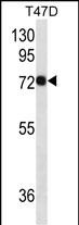
CD138 Antibody (Cat. #AM2157a) western blot analysis in T47D cell line lysates (35μg/lane).This demonstrates the CD138 antibody detected the CD138 protein (arrow).
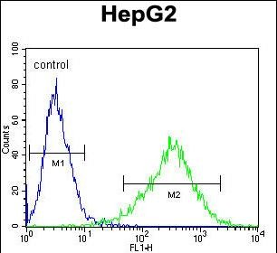
CD138 Antibody flow cytometric analysis of HepG2 cells (right histogram) compared to a negative control cell (left histogram).Alexa FluorR 488-conjugated donkey anti-mouse lgG secondary antibodies were used for the analysis
|
|
|
|
Confocal immunofluorescent analysis of CD138 antibody (Cat#AM2157a) with RPMI8266 cell (above) compared with Jurkat as negative cell line (below).followed by DyLight 488-conjugated goat anti-mouse lgG (H+L) Secondary Antibody (green).DAPI was used to stain the cell nucleus (blue).
|
|
|
| メーカー |
品番 |
包装 |
|
ABH
|
AM2157B
|
400 UL
|
※表示価格について
|

