|
※サムネイル画像をクリックすると拡大画像が表示されます。
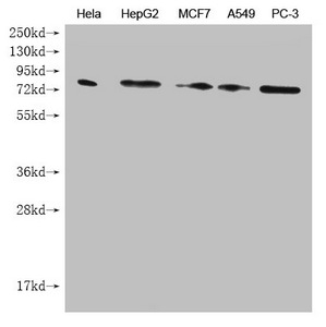
Western Blot
Positive WB detected in: Hela whole cell lysate, HepG2 whole cell lysate, MCF7 whole cell lysate, A549 whole cell lysate, PC-3 whole cell lysate
All lanes: SMURF1 antibody at 1:1000
Secondary
Goat polyclonal to mouse IgG at 1/50000 dilution
Predicted band size: 86 kDa
Observed band size: 86 KDa
Exposure time:5min
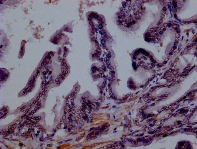
IHC image of CSB-MA875720A0m diluted at 1:100 and staining in paraffin-embedded human prostate tissue performed on a Leica BondTM system. After dewaxing and hydration, antigen retrieval was mediated by high pressure in a citrate buffer (pH 6.0). Section was blocked with 10% normal goat serum 30min at RT. Then primary antibody (1% BSA) was incubated at 4°C overnight. The primary is detected by a biotinylated secondary antibody and visualized using an HRP conjugated SP system.
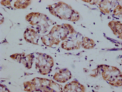
IHC image of CSB-MA875720A0m diluted at 1:100 and staining in paraffin-embedded human stomach tissue performed on a Leica BondTM system. After dewaxing and hydration, antigen retrieval was mediated by high pressure in a citrate buffer (pH 6.0). Section was blocked with 10% normal goat serum 30min at RT. Then primary antibody (1% BSA) was incubated at 4°C overnight. The primary is detected by a biotinylated secondary antibody and visualized using an HRP conjugated SP system.
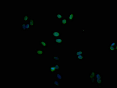
Immunofluorescence staining of Hela cells with CSB-MA875720A0m at 1:100, counter-stained with DAPI. The cells were fixed in 4% formaldehyde and blocked in 10% normal Goat Serum. The cells were incubated with the antibody overnight at 4°C. Nuclear DNA was labeled in blue with DAPI. The secondary antibody was FITC-conjugated AffiniPure Goat Anti-Mouse IgG (H+L).
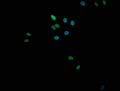
Immunofluorescence staining of HepG2 cells with CSB-MA875720A0m at 1:100, counter-stained with DAPI. The cells were fixed in 4% formaldehyde and blocked in 10% normal Goat Serum. The cells were incubated with the antibody overnight at 4°C. Nuclear DNA was labeled in blue with DAPI. The secondary antibody was FITC-conjugated AffiniPure Goat Anti-Mouse IgG (H+L).
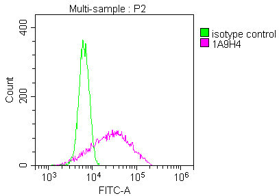
Overlay Peak curve showing Hela cells stained with CSB-MA875720A0m (red line) at 1:100. The cells were incubated in 10% normal goat serum to block non-specific protein-protein interactions followed by the antibody (1μg/1*106cells) for 1h at 4°C. The secondary antibody used was FITC-conjugated Goat Anti-Mouse IgG(H+L) at 1/100 dilution for 30min at 4°C. Isotype control antibody (green line) was mouse IgG1 (1μg/1*106cells) used under the same conditions. Acquisition of >10,000 events was performed.
|
|
|
|
Western Blot
Positive WB detected in: Hela whole cell lysate, HepG2 whole cell lysate, MCF7 whole cell lysate, A549 whole cell lysate, PC-3 whole cell lysate
All lanes: SMURF1 antibody at 1:1000
Secondary
Goat polyclonal to mouse IgG at 1/50000 dilution
Predicted band size: 86 kDa
Observed band size: 86 KDa
Exposure time:5min
|
|
| 別品名 |
E3 ubiquitin-protein ligase SMURF1 antibody; hSMURF1 antibody; KIAA1625 antibody; Smad specific E3 ubiquitin ligase 1 antibody; SMAD specific E3 ubiquitin protein ligase 1 antibody; Smad ubiquitination regulatory factor 1 antibody; SMAD-specific E3 ubiquitin-protein ligase 1 antibody; SMUF1_HUMAN antibody; SMURF 1 antibody; smurf1 antibody
|
| 種由来 |
Human
|
| 交差種 |
Human
|
| 適用 |
Western Blot
Enzyme Linked Immunosorbent Assay
Immunohistochemistry
Immuno Fluorescence
Flow Cytometry
|
| 免疫動物 |
Mouse
|
| クローン |
1A9H4
|
| 抗体クラス |
IgG2a
|
| 抗原部位 |
a.a.198-374
|
| 標識物 |
Unlabeled
|
| 精製度 |
Ig fraction - Protein G
|
| Accession No.(Gene/Protein) |
Q9HCE7
|
|
| メーカー |
品番 |
包装 |
|
CSB
|
CSB-MA875720A0M
|
100 UL
|
※表示価格について
| 当社在庫 |
なし
|
| 納期目安 |
2週間程度
|
| 保存温度 |
-20℃
|
|
※当社では商品情報の適切な管理に努めておりますが、表示される法規制情報は最新でない可能性があります。
また法規制情報の表示が無いものは、必ずしも法規制に非該当であることを示すものではありません。
商品のお届け前に最新の製品法規制情報をお求めの際はこちらへお問い合わせください。
|
※当社取り扱いの試薬・機器製品および受託サービス・創薬支援サービス(納品物、解析データ等)は、研究用としてのみ販売しております。
人や動物の医療用・臨床診断用・食品用としては、使用しないように、十分ご注意ください。
法規制欄に体外診断用医薬品と記載のものは除きます。
|
|
※リンク先での文献等のダウンロードに際しましては、掲載元の規約遵守をお願いします。
|
