
| 別品名 |
Antioxidant protein 1 antibody; AOP 1 antibody; AOP-1 antibody; AOP1 antibody; HBC189 antibody; MER5 antibody; MGC104387 antibody; MGC24293 antibody; mitochondrial antibody; peroxiredoxin 3 antibody; Peroxiredoxin III antibody; Peroxiredoxin-3 antibody; PRDX3 antibody; PRDX3_HUMAN antibody; PRO1748 antibody; Protein MER5 homolog antibody; PRX III antibody; Prx-III antibody; PRX3 antibody; SP 22 antibody; SP-22 antibody; SP22 antibody; Thioredoxin dependent peroxide reductase mitochondrial antibody; Thioredoxin-dependent peroxide reductase antibody
|
| 抗原部位 |
a.a.63-256
|
| 種由来 |
Human
|
| 標識物 |
Unlabeled
|
| 精製度 |
Ig fraction - Protein G
|
| 適用 |
Western Blot
Enzyme Linked Immunosorbent Assay
Immunohistochemistry
Immuno Fluorescence
Flow Cytometry
|
| 免疫動物 |
Mouse
|
| 抗体クラス |
IgG2b
|
| クローン |
12A10C12
|
| 交差種 |
Human
|
| Accession No.(Gene/Protein) |
P30048
|
| 形状 |
液状
|
|
※サムネイル画像をクリックすると拡大画像が表示されます。
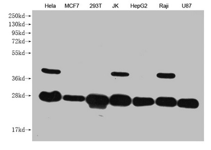
Western Blot
Positive WB detected in: Hela whole cell lysate, MCF7 whole cell lysate, 293T whole cell lysate, JK whole cell lysate, HepG2 whole cell lysate, Raji whole cell lysate, U87 whole cell lysate
All lanes: PRDX3 antibody at 1:1000
Secondary
Goat polyclonal to mouse IgG at 1/50000 dilution
Predicted band size: 28 kDa
Observed band size: 28 KDa
Exposure time:5min
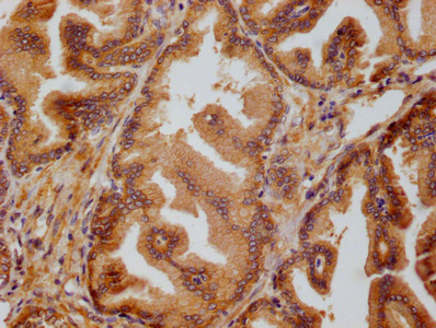
IHC image of CSB-MA018656A0m diluted at 1:50 and staining in paraffin-embedded human prostate tissue performed on a Leica BondTM system. After dewaxing and hydration, antigen retrieval was mediated by high pressure in a citrate buffer (pH 6.0). Section was blocked with 10% normal goat serum 30min at RT. Then primary antibody (1% BSA) was incubated at 4°C overnight. The primary is detected by a biotinylated secondary antibody and visualized using an HRP conjugated SP system.
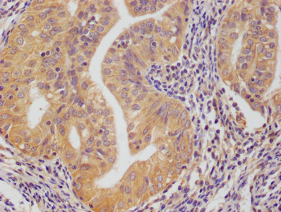
IHC image of CSB-MA018656A0m diluted at 1:50 and staining in paraffin-embedded human endometrial cance tissue performed on a Leica BondTM system. After dewaxing and hydration, antigen retrieval was mediated by high pressure in a citrate buffer (pH 6.0). Section was blocked with 10% normal goat serum 30min at RT. Then primary antibody (1% BSA) was incubated at 4°C overnight. The primary is detected by a biotinylated secondary antibody and visualized using an HRP conjugated SP system.
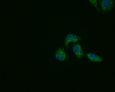
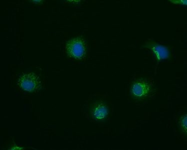
Immunofluorescence staining of Hela cells with(CSB-MA018656A0m)at 1:50, counter-stained with DAPI. The cells were fixed in 4% formaldehyde, permeabilized using 0.2% Triton X-100 and blocked in 10% normal Goat Serum. The cells were then incubated with the antibody overnight at 4°C. Nuclear DNA was labeled in blue with DAPI. The secondary antibody was FITC-conjugated AffiniPure Goat Anti-Mouse IgG (H+L).
Immunofluorescence staining of MCF7 cells with(CSB-MA018656A0m)at 1:50, counter-stained with DAPI. The cells were fixed in 4% formaldehyde, permeabilized using 0.2% Triton X-100 and blocked in 10% normal Goat Serum. The cells were then incubated with the antibody overnight at 4°C. Nuclear DNA was labeled in blue with DAPI. The secondary antibody was FITC-conjugated AffiniPure Goat Anti-Mouse IgG (H+L).
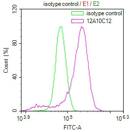
Overlay Peak curve showing Hela cells stained with CSB-MA018656A0m (red line) at 1:50. The cells were incubated in 10% normal goat serum to block non-specific protein-protein interactions followed by the antibody (1μg/1*106cells) for 1h at 4°C. The secondary antibody used was FITC-conjugated Goat Anti-Mouse IgG(H+L) at 1/100 dilution for 30min at 4°C. Isotype control antibody (green line) was mouse IgG1 (1μg/1*106cells) used under the same conditions. Acquisition of >10,000 events was performed.
|
|
|
|
Western Blot
Positive WB detected in: Hela whole cell lysate, MCF7 whole cell lysate, 293T whole cell lysate, JK whole cell lysate, HepG2 whole cell lysate, Raji whole cell lysate, U87 whole cell lysate
All lanes: PRDX3 antibody at 1:1000
Secondary
Goat polyclonal to mouse IgG at 1/50000 dilution
Predicted band size: 28 kDa
Observed band size: 28 KDa
Exposure time:5min
|
|
|
| メーカー |
品番 |
包装 |
|
CSB
|
CSB-MA018656A0M
|
50 UL
|
※表示価格について
| 当社在庫 |
なし
|
| 納期目安 |
2週間程度
|
| 保存温度 |
-20℃
|
|

