
| 別品名 |
Antigen CD48 antibody; B cell membrane protein antibody; B lymphocyte activation marker BLAST 1 antibody; B-cell activation marker antibody; B-lymphocyte activation marker BLAST-1 antibody; BCM 1 surface antigen antibody; BCM1 antibody; BCM1 surface antigen antibody; BLAST 1 antibody; BLAST antibody; BLAST1 antibody; CD 48 antibody; CD48 antibody; CD48 antigen (B cell membrane protein) antibody; CD48 antigen antibody; CD48 molecule antibody; CD48 protein antibody; CD48_HUMAN antibody; hCD48 antibody; Leucocyte antigen MEM 102 antibody; Leukocyte antigen MEM-102 antibody; mCD48 antibody; MEM 102 antibody; MEM-102 antibody; MEM102 antibody; Signaling lymphocytic activation molecule 2 antibody; SLAM family member 2 antibody; SLAMF 2 antibody; SLAMF2 antibody; TCT.1 antibody
|
| 抗原部位 |
a.a.27-220
|
| 種由来 |
Human
|
| 標識物 |
Unlabeled
|
| 精製度 |
Ig fraction - Protein G
|
| 適用 |
Western Blot
Enzyme Linked Immunosorbent Assay
Immuno Fluorescence
Flow Cytometry
|
| 免疫動物 |
Mouse
|
| 抗体クラス |
IgG1
|
| クローン |
6F9H2
|
| 交差種 |
Human
Mouse
|
| Accession No.(Gene/Protein) |
P09326
|
| 形状 |
液状
|
|
※サムネイル画像をクリックすると拡大画像が表示されます。
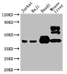
Western Blot
Positive WB detected in: Jurkat whole cell lysate, Raji whole cell lysate, Daudi whole cell lysate, Mouse liver tissue
All lanes: CD48 antibody at 1:4000
Secondary
Goat polyclonal to mouse IgG at 1/50000 dilution
Predicted band size: 28, 20KDa
Observed band size: 43 KDa
Exposure time:5min
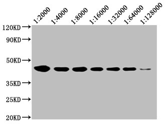
Western Blot
Positive WB detected in: 20ug Raji whole cell lysate
CD48 antibody at 1:2000, 1:4000, 1:8000, 1:16000, 1:32000, 1:64000, 1:128000
Secondary
Goat polyclonal to mouse IgG at 1/50000 dilution
Predicted band size: 28, 20 KDa
Observed band size: 43 KDa
Exposure time:5min
Immunofluorescence staining of JK cells with CSB-MA004941A0m at 1:100, counter-stained with DAPI. The cells were fixed in 4% formaldehyde and blocked in 10% normal Goat Serum. The cells were incubated with the antibody overnight at 4°C. Nuclear DNA was labeled in blue with DAPI. The secondary antibody was FITC-conjugated AffiniPure Goat Anti-Mouse IgG (H+L).

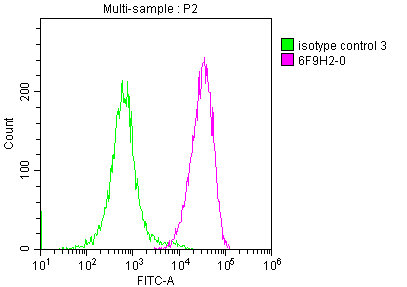
Overlay Peak curve showing JK cells stained with CSB-MA004941A0m (red line) at 1:200. The cells were incubated in 10% normal goat serum to block non-specific protein-protein interactions followed by the antibody (1μg/1*106 cells) for 1h at 4°C. The secondary antibody used was FITC-conjugated Goat Anti-Mouse IgG(H+L) at 1/100 dilution for 30min at 4°C. Isotype control antibody (green line) was mouse IgG1 (1μg/1*106 cells) used under the same conditions. Acquisition of >10,000 events was performed.
|
|
|
|
Western Blot
Positive WB detected in: Jurkat whole cell lysate, Raji whole cell lysate, Daudi whole cell lysate, Mouse liver tissue
All lanes: CD48 antibody at 1:4000
Secondary
Goat polyclonal to mouse IgG at 1/50000 dilution
Predicted band size: 28, 20KDa
Observed band size: 43 KDa
Exposure time:5min
|
|
|
| メーカー |
品番 |
包装 |
|
CSB
|
CSB-MA004941A0M
|
50 UL
|
※表示価格について
| 当社在庫 |
なし
|
| 納期目安 |
2週間程度
|
| 保存温度 |
-20℃
|
|

