
| 別品名 |
Histone-binding protein RBBP4 (Chromatin assembly factor 1 subunit C) (CAF-1 subunit C) (Chromatin assembly factor I p48 subunit) (CAF-I 48 kDa subunit) (CAF-I p48) (Nucleosome-remodeling factor subunit RBAP48) (Retinoblastoma-binding protein 4) (RBBP-4) (Retinoblastoma-binding protein p48), RBBP4, RBAP48
|
| 種由来 |
Human
|
| 標識物 |
Unlabeled
|
| 精製度 |
Affinity Purified
|
| 適用 |
Western Blot
Enzyme Linked Immunosorbent Assay
Immunohistochemistry
Immuno Fluorescence
|
| 免疫動物 |
Rabbit Mono
|
| 抗体クラス |
IgG
|
| クローン |
3E9
|
| 交差種 |
Human
Mouse
Rat
|
| Accession No.(Gene/Protein) |
Q09028
|
| 形状 |
液状
|
|
※サムネイル画像をクリックすると拡大画像が表示されます。
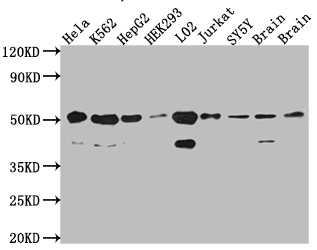
Western Blot
Positive WB detected in: Hela whole cell lysate, K562 whole cell lysate, HepG2 whole cell lysate, HEK293 whole cell lysate, L02 whole cell lysate, Jurkat whole cell lysate, SH-SY5Y whole cell lysate, Mouse Brain whole cell lysate, Rat Brain cell lysate
All lanes: RbAp48 antibody at 1:1000
Secondary
Goat polyclonal to rabbit IgG at 1/50000 dilution
Predicted band size: 48, 48, 47, 44 kDa
Observed band size: 53, 40 kDa
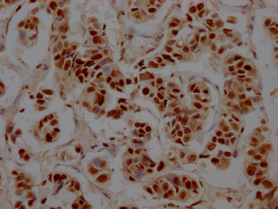
IHC image of CSB-RA915915A0HU diluted at 1:100 and staining in paraffin-embedded human breast cancer performed on a Leica BondTM system. After dewaxing and hydration, antigen retrieval was mediated by high pressure in a citrate buffer (pH 6.0). Section was blocked with 10% normal goat serum 30min at RT. Then primary antibody (1% BSA) was incubated at 4℃ overnight. The primary is detected by a Goat anti-rabbit IgG polymer labeled by HRP and visualized using 0.05% DAB.
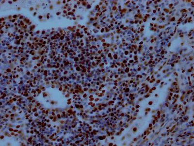
IHC image of CSB-RA915915A0HU diluted at 1:100 and staining in paraffin-embedded human lung tissue performed on a Leica BondTM system. After dewaxing and hydration, antigen retrieval was mediated by high pressure in a citrate buffer (pH 6.0). Section was blocked with 10% normal goat serum 30min at RT. Then primary antibody (1% BSA) was incubated at 4℃ overnight. The primary is detected by a Goat anti-rabbit IgG polymer labeled by HRP and visualized using 0.05% DAB.
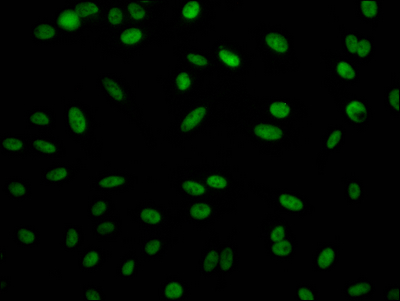
Immunofluorescence staining of Hela Cells with CSB-RA915915A0HU at 1:50, counter-stained with DAPI. The cells were fixed in 4% formaldehyde, permeated by 0.2% TritonX-100, and blocked in 10% normal Goat Serum. The cells were then incubated with the antibody overnight at 4℃. Nuclear DNA was labeled in blue with DAPI. The secondary antibody was FITC-conjugated AffiniPure Goat Anti-Rabbit IgG (H+L).
|
|
|
|
Western Blot
Positive WB detected in: Hela whole cell lysate, K562 whole cell lysate, HepG2 whole cell lysate, HEK293 whole cell lysate, L02 whole cell lysate, Jurkat whole cell lysate, SH-SY5Y whole cell lysate, Mouse Brain whole cell lysate, Rat Brain cell lysate
All lanes: RbAp48 antibody at 1:1000
Secondary
Goat polyclonal to rabbit IgG at 1/50000 dilution
Predicted band size: 48, 48, 47, 44 kDa
Observed band size: 53, 40 kDa
|
|
|
| メーカー |
品番 |
包装 |
|
CSB
|
CSB-RA915915A0HU
|
50 UL
|
※表示価格について
| 当社在庫 |
なし
|
| 納期目安 |
2週間程度
|
| 保存温度 |
-20℃
|
|

