
| 別品名 |
Tissue factor (TF) (Coagulation factor III) (Thromboplastin) (CD antigen CD142), F3
|
| 種由来 |
Human
|
| 標識物 |
Unlabeled
|
| 精製度 |
Affinity Purified
|
| 適用 |
Western Blot
Enzyme Linked Immunosorbent Assay
Immunohistochemistry
|
| 免疫動物 |
Rabbit Mono
|
| 抗体クラス |
IgG
|
| クローン |
8B10
|
| 交差種 |
Human
Rat
|
| Accession No.(Gene/Protein) |
P13726
|
| 形状 |
液状
|
|
※サムネイル画像をクリックすると拡大画像が表示されます。
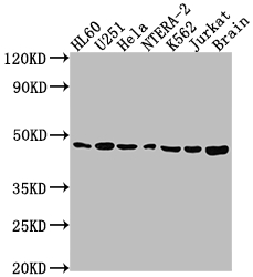
Western Blot
Positive WB detected in: HL60 whole cell lysate, U251 whole cell lysate, Hela whole cell lysate, NTERA-2 whole cell lysate, K562 whole cell lysate, Jurkat whole cell lysate, Rat brain tissue
All lanes: F3 antibody at 1:1500
Secondary
Goat polyclonal to rabbit IgG at 1/50000 dilution
Predicted band size: 34, 28 kDa
Observed band size: 46 kDa
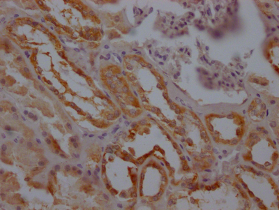
IHC image of CSB-RA776663A0HU diluted at 1:100 and staining in paraffin-embedded human kidney tissue performed on a Leica BondTM system. After dewaxing and hydration, antigen retrieval was mediated by high pressure in a citrate buffer (pH 6.0). Section was blocked with 10% normal goat serum 30min at RT. Then primary antibody (1% BSA) was incubated at 4℃ overnight. The primary is detected by a Goat anti-rabbit IgG polymer labeled by HRP and visualized using 0.05% DAB.
|
|
|
|
Western Blot
Positive WB detected in: HL60 whole cell lysate, U251 whole cell lysate, Hela whole cell lysate, NTERA-2 whole cell lysate, K562 whole cell lysate, Jurkat whole cell lysate, Rat brain tissue
All lanes: F3 antibody at 1:1500
Secondary
Goat polyclonal to rabbit IgG at 1/50000 dilution
Predicted band size: 34, 28 kDa
Observed band size: 46 kDa
|
|
|
| メーカー |
品番 |
包装 |
|
CSB
|
CSB-RA776663A0HU
|
100 UL
|
※表示価格について
| 当社在庫 |
なし
|
| 納期目安 |
2週間程度
|
| 保存温度 |
-20℃
|
|

