|
※サムネイル画像をクリックすると拡大画像が表示されます。
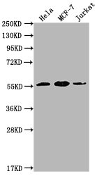
Western Blot
Positive WB detected in: Hela whole cell lysate, MCF-7 whole cell lysate, Jurkat whole cell lysate
All lanes: PKM antibody at 1:1000
Secondary
Goat polyclonal to Mouse IgG at 1/10000 dilution
Predicted band size: 58 kDa
Observed band size: 58 KDa
Exposure time: 1min
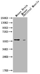
Western Blot
Positive WB detected in: Mouse Brain tissue, Mouse Skeldtal Muscle tissue
All lanes: PKM antibody at 1:1000
Secondary
Goat polyclonal to Mouse IgG at 1/10000 dilution
Predicted band size: 58 kDa
Observed band size: 58 KDa
Exposure time: 5min
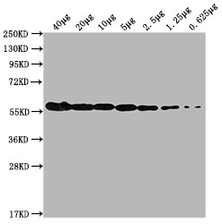
Western Blot
Positive WB detected in: MCF-7 whole cell lysate at 40μg, 20μg, 10μg, 5μg, 2.5μg, 1.25μg, 0.625μg
All lanes: PKM antibody at 1:1000
Secondary
Goat polyclonal to Mouse IgG at 1/10000 dilution
Predicted band size: 58 kDa
Observed band size: 58 KDa
Exposure time: 5min
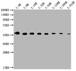
Western Blot
Positive WB detected in: MCF-7 whole cell lysate
All lanes: PKM antibody at 1:4000, 1:8000, 1:16000, 1:32000, 1:64000, 1:128000, 1:256000, 1:512000
Secondary
Goat polyclonal to Mouse IgG at 1/10000 dilution
Predicted band size: 58 kDa
Observed band size: 58 KDa
Exposure time: 5min
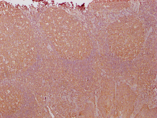
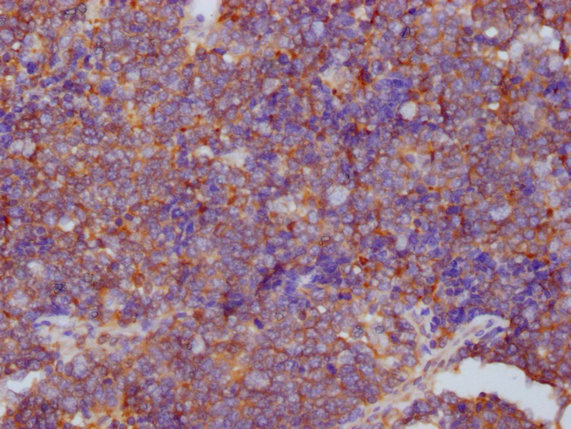
IHC image of CSB-MA018072A1m diluted at 1:1000 and staining in paraffin-embedded human tonsil tissue performed on a Leica BondTM system. After dewaxing and hydration, antigen retrieval was mediated by high pressure in a citrate buffer (pH 6.0). Section was blocked with 10% normal goat serum 30min at RT. Then primary antibody (1% BSA) was incubated at 4°C overnight. The primary is detected by a biotinylated secondary antibody and visualized using an HRP conjugated SP system.
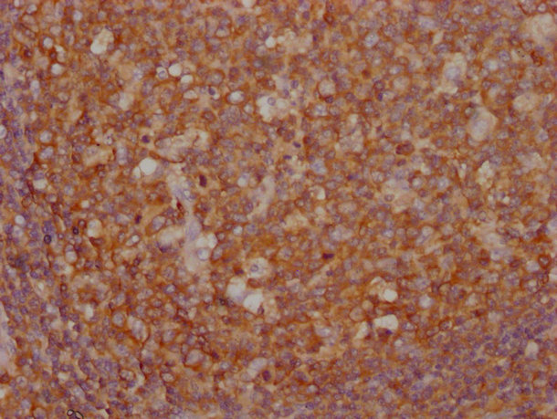
IHC image of CSB-MA018072A1m diluted at 1:1000 and staining in paraffin-embedded human tonsil tissue performed on a Leica BondTM system. After dewaxing and hydration, antigen retrieval was mediated by high pressure in a citrate buffer (pH 6.0). Section was blocked with 10% normal goat serum 30min at RT. Then primary antibody (1% BSA) was incubated at 4°C overnight. The primary is detected by a biotinylated secondary antibody and visualized using an HRP conjugated SP system.
IHC image of CSB-MA018072A1m diluted at 1:1000 and staining in paraffin-embedded human tonsil tissue performed on a Leica BondTM system. After dewaxing and hydration, antigen retrieval was mediated by high pressure in a citrate buffer (pH 6.0). Section was blocked with 10% normal goat serum 30min at RT. Then primary antibody (1% BSA) was incubated at 4°C overnight. The primary is detected by a biotinylated secondary antibody and visualized using an HRP conjugated SP system.
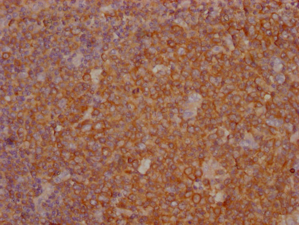
IHC image of CSB-MA018072A1m diluted at 1:1000 and staining in paraffin-embedded human lung cancer tissue performed on a Leica BondTM system. After dewaxing and hydration, antigen retrieval was mediated by high pressure in a citrate buffer (pH 6.0). Section was blocked with 10% normal goat serum 30min at RT. Then primary antibody (1% BSA) was incubated at 4°C overnight. The primary is detected by a biotinylated secondary antibody and visualized using an HRP conjugated SP system.
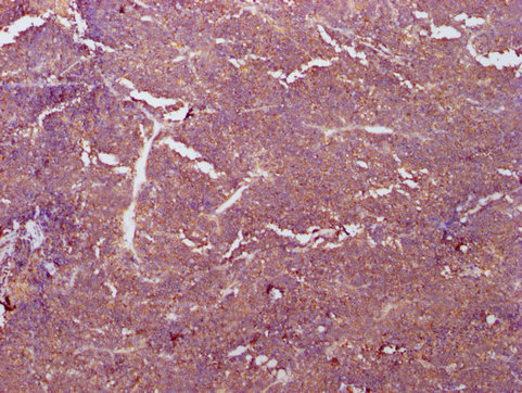
IHC image of CSB-MA018072A1m diluted at 1:1000 and staining in paraffin-embedded human lung cancer tissue performed on a Leica BondTM system. After dewaxing and hydration, antigen retrieval was mediated by high pressure in a citrate buffer (pH 6.0). Section was blocked with 10% normal goat serum 30min at RT. Then primary antibody (1% BSA) was incubated at 4°C overnight. The primary is detected by a biotinylated secondary antibody and visualized using an HRP conjugated SP system.
|

