|
※サムネイル画像をクリックすると拡大画像が表示されます。
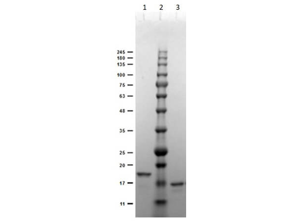
SDS-PAGE Results of Recombinant Anti-DIG (VhH) Single Domain Antibody. Lane 1: Anti-DIG-VhH (Clone DIG44) Reduced (1μg). Lane 2: Opal Prestained Molecular Weight Marker (p/n MB-210-0500). Lane 3: Anti-DIG-VhH (Clone DIG44) Non-Reduced (1μg). 4-20% Gel Coomassie Blue stained. Predicted MW: ~15kDa. Observed MW: ~18kDa Reduced, ~16kDa Non-Reduced.
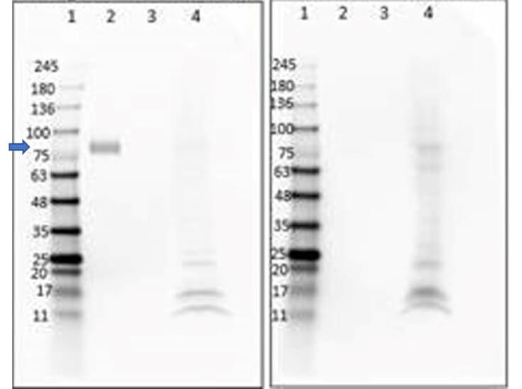
Western Blot of Anti-DIG Single Domain Antibody. Lane 1: Opal Prestained Molecular Weight Marker (p/n MB-210-0500) (5μL). Lane 2: DIG-BSA conjugated (0.2μg). Lane 3: BSA only (0.2μg). Lane 4: HEK293T lysate (p/n W09-001-GX5) (20μg). Left Blot - Primary Antibody: Anti-DIG (VhH) at 1μg/mL incubated overnight at 4°C. Right Blot - No Primary. Secondary Only Control. Secondary Antibody: Mouse Anti-6X HIS DyLight?649 (p/n 200-343-382) at 1:1000 incubated for 1hr at RT. Blocking buffer: Universal Buffer (p/n MB-070) for 2hrs at RT. Expected MW: ~80kDa in DIG+BSA. Observed MW: (arrow) ~80kDa in DIG+BSA; secondary background seen in current tested lot of HEK293T Lysate.
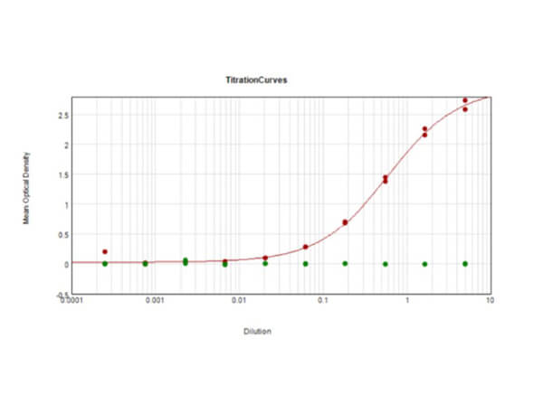
ELISA Results of purified recombinant Anti-DIG Single Domain Antibody reactive against BSA-conjugated DIG. The working dilution for clone DIG44 is 1:1000. Each well was coated in duplicate with 1.0μg BSA-conjugated DIG (Red) and BSA only (Green). The starting dilution of antibody was 45μg/ml and the X-axis represents the Log10 of a 3-fold dilution. This titration is a 4-parameter curve fit where the IC50 is defined as the titer of the antibody. Assay performed using Sodium Carbonate blocking buffer, Mouse Anti-6X HIS Epitope Tag HRP conjugated Antibody (p/n 200-303-382) and TMB substrate (p/n TMBE-1000).
|
|
|
|
SDS-PAGE Results of Recombinant Anti-DIG (VhH) Single Domain Antibody. Lane 1: Anti-DIG-VhH (Clone DIG44) Reduced (1μg). Lane 2: Opal Prestained Molecular Weight Marker (p/n MB-210-0500). Lane 3: Anti-DIG-VhH (Clone DIG44) Non-Reduced (1μg). 4-20% Gel Coomassie Blue stained. Predicted MW: ~15kDa. Observed MW: ~18kDa Reduced, ~16kDa Non-Reduced.
|
|
| 別品名 |
anti-Digoxigenin, Digoxigenin (DIG), 1672-46-4, Lanadigenin, Digoxigenine, UNII-NQ1SX9LNAU, DIG, CHEBI:42098, HSDB 7108, NQ1SX9LNAU, EINECS 216-806-2, BRN 0096479, 4-(3,12,14-Trihydroxy-10,13-Dimethyl-Hexadecahydro-Cyclopenta[a]Phenanthren-17-Yl)-5h-Furan-2-One
|
| 種由来 |
Escherichia coli
|
交差種以外の
交差情報
(微交差など) |
[Reactivity]Digitalis sp.
|
| 適用 |
Western Blot
Enzyme Linked Immunosorbent Assay
|
| クローン |
DIG44
|
| 標識物 |
Unlabeled
|
| Gene Symbol |
Digoxigenin
|
| タグ(タンパク質) |
6X HIS
|
| 発現系 |
大腸菌
|
|
| メーカー |
品番 |
包装 |
|
RKL
|
400-001-MG3
|
50 UG
|
※表示価格について
|
※当社では商品情報の適切な管理に努めておりますが、表示される法規制情報は最新でない可能性があります。
また法規制情報の表示が無いものは、必ずしも法規制に非該当であることを示すものではありません。
商品のお届け前に最新の製品法規制情報をお求めの際はこちらへお問い合わせください。
|
※当社取り扱いの試薬・機器製品および受託サービス・創薬支援サービス(納品物、解析データ等)は、研究用としてのみ販売しております。
人や動物の医療用・臨床診断用・食品用としては、使用しないように、十分ご注意ください。
法規制欄に体外診断用医薬品と記載のものは除きます。
|
|
※リンク先での文献等のダウンロードに際しましては、掲載元の規約遵守をお願いします。
|
|
※CAS Registry Numbers have not been verified by CAS and may be inaccurate.
|
