|
※サムネイル画像をクリックすると拡大画像が表示されます。
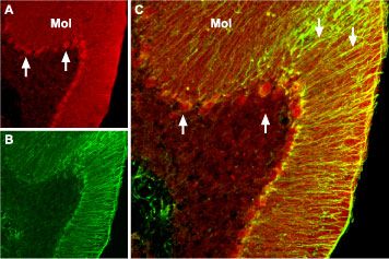
Expression of BDNF in mouse cerebellum - Immunohistochemical staining of mouse cerebellum with?Anti-BDNF?Antibody (#ANT-010). A. BDNF (red) appears in Purkinje cells (upward pointing arrows) and is distributed diffusely in the molecular layer (Mol) including in astrocytic fibers (downward pointing arrows). B. Staining of astrocytic fibers with glial fibrillary acidic protein (green) in the same section demonstrates the distribution of BDNF to neuronal as well as to astrocytic cellular components. C. Confocal merge of BDNF and GFAP.
|
|
|
|
Expression of BDNF in mouse cerebellum - Immunohistochemical staining of mouse cerebellum with?Anti-BDNF?Antibody (#ANT-010). A. BDNF (red) appears in Purkinje cells (upward pointing arrows) and is distributed diffusely in the molecular layer (Mol) including in astrocytic fibers (downward pointing arrows). B. Staining of astrocytic fibers with glial fibrillary acidic protein (green) in the same section demonstrates the distribution of BDNF to neuronal as well as to astrocytic cellular components. C. Confocal merge of BDNF and GFAP.
|
|
| 概要 |
A Blocking Peptide for Anti-BDNF Antibody
|
| [注意事項] |
抗体と専用ブロッキングペプチドを同時購入頂く場合のみ、専用ブロッキングペプチドが40%OFFとなります。同時購入には注文書の他に専用申込書が必要です。
※専用ブロッキングペプチドの品番は、メーカーWEBページ右欄の「Blocking Peptide Control」や「Specific Control Products」(ピンク字表示)に記載されています。
|
|
| メーカー |
品番 |
包装 |
|
ALO
|
BLP-NT010
|
40 UG
|
※表示価格について
| 当社在庫 |
なし
|
| 納期目安 |
約10日
|
| 保存温度 |
-20℃
|
|
※当社では商品情報の適切な管理に努めておりますが、表示される法規制情報は最新でない可能性があります。
また法規制情報の表示が無いものは、必ずしも法規制に非該当であることを示すものではありません。
商品のお届け前に最新の製品法規制情報をお求めの際はこちらへお問い合わせください。
|
※当社取り扱いの試薬・機器製品および受託サービス・創薬支援サービス(納品物、解析データ等)は、研究用としてのみ販売しております。
人や動物の医療用・臨床診断用・食品用としては、使用しないように、十分ご注意ください。
法規制欄に体外診断用医薬品と記載のものは除きます。
|
|
※リンク先での文献等のダウンロードに際しましては、掲載元の規約遵守をお願いします。
|
|
※CAS Registry Numbers have not been verified by CAS and may be inaccurate.
|
