
| 別品名 |
HTPHLP antibody; IAP associated factor VIAF1 antibody; MGC3062 antibody; PDCL 3 antibody; Pdcl3 antibody; PDCL3_HUMAN antibody; PHLP2A antibody; PHLP3 antibody; Phosducin like 3 antibody; Phosducin like protein 3 antibody; Phosducin-like protein 3 antibody; PhPL3 antibody; VIAF 1 antibody; VIAF antibody; VIAF-1 antibody; VIAF1 antibody; Viral IAP associated factor 1 antibody; Viral IAP-associated factor 1 antibody
|
| 抗原部位 |
a.a.1-239
|
| 種由来 |
Human
|
| 標識物 |
Unlabeled
|
| 精製度 |
Ig fraction - Protein G
|
| 適用 |
Western Blot
Enzyme Linked Immunosorbent Assay
Immuno Fluorescence
|
| 免疫動物 |
Rabbit
|
| 抗体クラス |
IgG
|
| 交差種 |
Human
|
| Accession No.(Gene/Protein) |
Q9H2J4
|
| 形状 |
液状
|
|
※サムネイル画像をクリックすると拡大画像が表示されます。
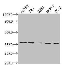
Western Blot
Positive WB detected in: A2780 whole cell lysate, 293 whole cell lysate, U251 whole cell lysate, MCF-7 whole cell lysate, PC-3 whole cell lysate
All lanes: PDCL3 antibody at 1:2000
Secondary
Goat polyclonal to rabbit IgG at 1/50000 dilution
Predicted band size: 28 kDa
Observed band size: 37 kDa
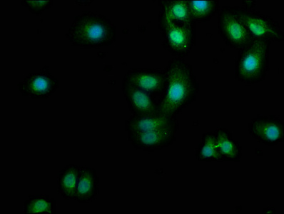
Immunofluorescence staining of HepG2 cells with CSB-PA872479LA01HU at 1:100, counter-stained with DAPI. The cells were fixed in 4% formaldehyde, permeabilized using 0.2% Triton X-100 and blocked in 10% normal Goat Serum. The cells were then incubated with the antibody overnight at 4°C. The secondary antibody was Alexa Fluor 488-congugated AffiniPure Goat Anti-Rabbit IgG(H+L).
|
|
|
|
Western Blot
Positive WB detected in: A2780 whole cell lysate, 293 whole cell lysate, U251 whole cell lysate, MCF-7 whole cell lysate, PC-3 whole cell lysate
All lanes: PDCL3 antibody at 1:2000
Secondary
Goat polyclonal to rabbit IgG at 1/50000 dilution
Predicted band size: 28 kDa
Observed band size: 37 kDa
|
|
|
| メーカー |
品番 |
包装 |
|
CSB
|
CSB-PA872479LA01HU
|
50 UL
|
※表示価格について
| 当社在庫 |
なし
|
| 納期目安 |
2週間程度
|
| 保存温度 |
-20℃
|
|

