|
※サムネイル画像をクリックすると拡大画像が表示されます。
Western blot analysis of extracts of various cell lines, using TOM20 antibody (16-790) at 1:1000 dilution.
Secondary antibody: HRP Goat Anti-Rabbit IgG (H+L) at 1:10000 dilution.
Lysates/proteins: 25ug per lane.
Blocking buffer: 3% nonfat dry milk in TBST.
Detection: ECL Basic Kit.
Exposure time: 5min.

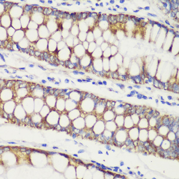
Immunohistochemistry of paraffin-embedded human colon carcinoma using TOM20 antibody (16-790) at dilution of 1:100 (40x lens).
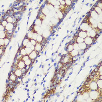
Immunohistochemistry of paraffin-embedded human colon using TOM20 antibody (16-790) at dilution of 1:100 (40x lens).
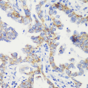
Immunohistochemistry of paraffin-embedded human lung cancer using TOM20 antibody (16-790) at dilution of 1:100 (40x lens).
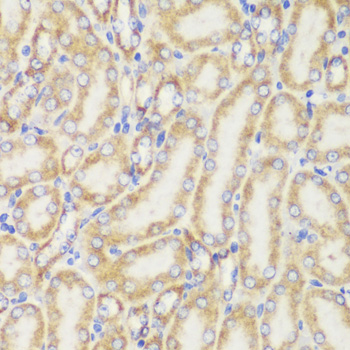
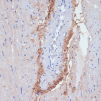
Immunohistochemistry of paraffin-embedded mouse kidney using TOM20 antibody (16-790) at dilution of 1:100 (40x lens).
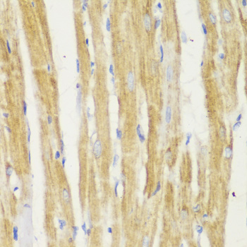
Immunohistochemistry of paraffin-embedded rat heart using TOM20 antibody (16-790) at dilution of 1:100 (40x lens).
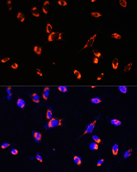
Immunofluorescence analysis of C6 cells using TOM20 antibody (16-790) at dilution of 1:100. Blue: DAPI for nuclear staining.
Immunofluorescence analysis of NIH/3T3 using TOM20 antibody (16-790) at dilution of 1:100. Blue: DAPI for nuclear staining.
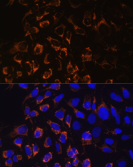
Immunofluorescence analysis of U2OS cells using TOM20 antibody (16-790) at dilution of 1:100. Blue: DAPI for nuclear staining.
|

