
| 別品名 |
Rabbit Anti-Microtubule Associated Protein 1 Light Chain 3 Beta Antibody, Rabbit Anti-LC3B Antibody, Microtubule-Associated Proteins 1A/1B Light Chain 3B, Autophagy-Related Ubiquitin-Like Modifier LC3 B, MAP1 Light Chain 3-Like Protein 2, MAP1A/MAP1B Light Chain 3 B, MAP1A/MAP1B LC3 B, Microtubule-Associated Protein 1 Light Chain 3 Beta, Autophagy-Related Protein LC3 B, MAP1A/1BLC3, MAP1LC3B-A, MAP1ALC3, ATG8F, LC3B
|
| 抗原部位 |
Internal
|
| 種由来 |
Human
|
| 標識物 |
Unlabeled
|
| 精製度 |
Affinity Purified
|
| 適用 |
Western Blot
Enzyme Linked Immunosorbent Assay
Immunohistochemistry
Flow Cytometry
|
| 免疫動物 |
Rabbit
|
| 交差種 |
Human
|
| GENE ID |
81631
|
| Accession No.(Gene/Protein) |
NP_073729, Q9GZQ8
|
| Gene Symbol |
MAP1LC3B
|
| 形状 |
液状
|
| その他 |
[Buffer]0.02 M Potassium Phosphate, 0.15 M Sodium Chloride, pH 7.2
|
| [注意事項] |
濃度はロットによって異なる可能性があります。メーカーDS及びCoAからご確認ください。
|
|
※サムネイル画像をクリックすると拡大画像が表示されます。
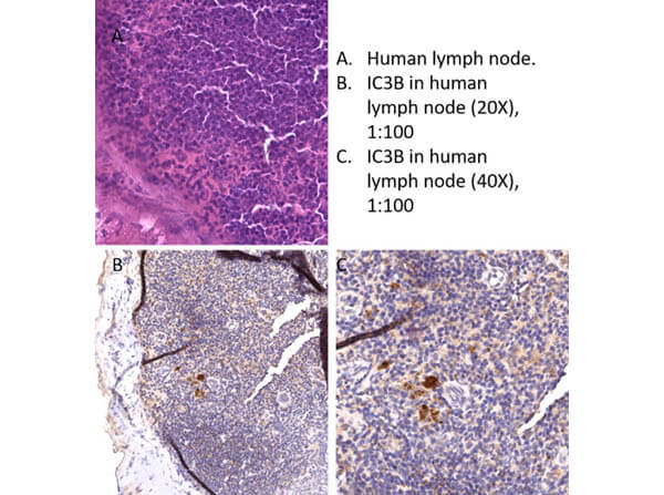
Immunohistochemistry of Rabbit Anti LC3B Antibody. Tissue: human lymph node. Fixative: none. Antigen Retrieval: HIER using Cirate Buffer for 20 minutes. Primary Antibody: Anti LC3B antibody at 1:100 at RT for 30 minutes. Secondary Antibody: Anti Rabbit Poly HRP IgG. Ready to Use at RT for 8 minutes. Counterstain: Hematoxylin. Substrate: DAB. Results: This antibody showed staining throughout most of the lymph node with focal strong staining. Focal cell staining strongly in the lymph node is the pattern expected.
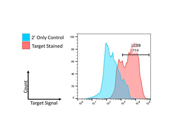
Flow Cytometry of Rabbit Anti-LC3B Antibody. Cells: U251-MG cells. Primary Antibody: Anti-LC3B at 2.5μg/mL in 100μL FACS buffer for 30 minutes at RT. Secondary Antibody: Donkey anti-Rabbit IgG DyLight?488 (p/n 611-741-127) at 2.5μg/mL in 100μL FACS buffer for 30 minutes at RT. Buffer: FACS/IF buffer (p/n MB-086-0500). Analysis: Distinct positive shifts in fluorescence have been observed in all samples tested following permeabilization of the cell sample. This is indicative of specificity and affinity of each antibody toward its target.
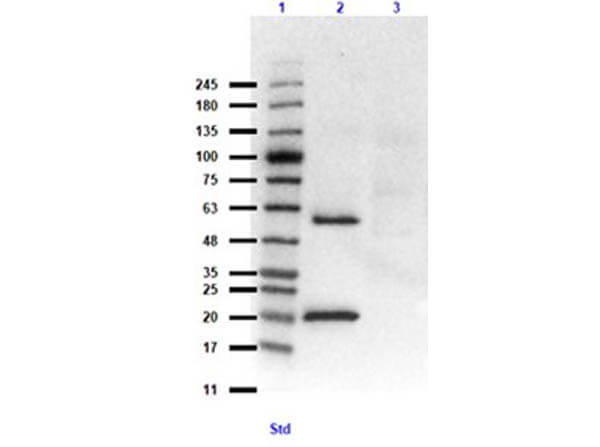
Western Blot of Rabbit Anti-LC3B Antibody. Lane 1: Opal Prestained Molecular Weight Ladder (p/n MB-210-0500). Lane 2: MAP1LC3B overexpressing HEK293 (10ug) [+]. Lane 3: HEK293T lysate (p/n W09-001-GX5) (10 ug) [-]. Primary Antibody: Anti-LC3B Antibody at 1:1000 overnight at 2-8C. Secondary Antibody: Goat anti-Rabbit IgG HRP (p/n 611-103-122) at 1:70,000 for 30 minutes. Block: BlockOut Buffer (p/n MB-073). Exposure: 15 sec. Predicted MW: ~14.6, ~63kDa for overexpressing lysates. Observed MW: ~20, 60kDa.
|
|
|
|
Immunohistochemistry of Rabbit Anti LC3B Antibody. Tissue: human lymph node. Fixative: none. Antigen Retrieval: HIER using Cirate Buffer for 20 minutes. Primary Antibody: Anti LC3B antibody at 1:100 at RT for 30 minutes. Secondary Antibody: Anti Rabbit Poly HRP IgG. Ready to Use at RT for 8 minutes. Counterstain: Hematoxylin. Substrate: DAB. Results: This antibody showed staining throughout most of the lymph node with focal strong staining. Focal cell staining strongly in the lymph node is the pattern expected.
|
|
|
| メーカー |
品番 |
包装 |
|
RKL
|
600-401-MN5S
|
25 UL
|
※表示価格について
| 当社在庫 |
なし
|
| 納期目安 |
約10日
|
| 保存温度 |
-20℃
|
|

