| 別品名 |
Rabbit Anti-Beclin-1, Beclin1 Antibody, Beclin 1 Antibody, BECN1, Coiled-Coil Myosin-Like BCL2-Interacting Protein,?Beclin 1 Autophagy Related, Beclin 1 (Coiled-Coil, Moesin-Like BCL2 Interacting Protein),?ATG6 Autophagy Related 6 Homolog, Protein GT197, VPS30, GT197
|
| 抗原部位 |
N-Terminus
|
| 種由来 |
Human
|
| 標識物 |
Unlabeled
|
| 精製度 |
Affinity Purified
|
| 適用 |
Western Blot
Enzyme Linked Immunosorbent Assay
Immunohistochemistry
Immuno Fluorescence
|
| 免疫動物 |
Rabbit
|
| 交差種 |
Human
Mouse
|
| GENE ID |
8678
|
| Accession No.(Gene/Protein) |
NP_001300927, Q14457
|
| Gene Symbol |
BECN1
|
| 形状 |
液状
|
| その他 |
[Buffer]0.02 M Potassium Phosphate, 0.15 M Sodium Chloride, pH 7.2
|
| [注意事項] |
濃度はロットによって異なる可能性があります。メーカーDS及びCoAからご確認ください。
|
|
※サムネイル画像をクリックすると拡大画像が表示されます。
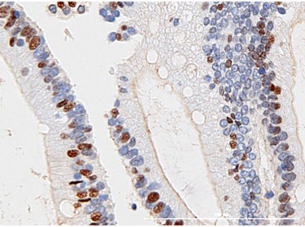
Immunohistochemistry of Rabbit Anti Beclin 1 Antibody. Tissue: Human colon cancer tissue (40X). Fixative: none. Antigen Retrieval: Heat induced epitope retrieval (HIER). Primary Antibody: Anti Beclin 1 at 1:100 for 30mins at RT. Secondary Antibody: Anti Rabbit Poly HRP IgG ready to use 8 min at RT. Counterstain: hematoxylin. Expected Localization: nuclear or cytoplasmic staining of normal cells. Less staining of cancer cells. Result Analysis: Passed. Epithelial layer staining, nuclear staining, cell cell junction staining.

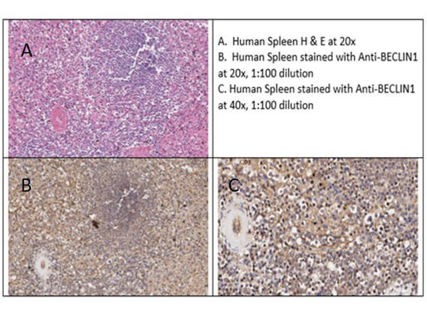
Immunohistochemistry of Rabbit Anti-Beclin 1 Antibody. Tissue: Human Spleen. Fixative: none. Antigen Retrieval: HIER using Citrate buffer for 20 minutes. Primary Antibody: Anti-Beclin 1 at 1:100 for 30mins at RT. Secondary Antibody: Anti-Rabbit Poly-HRP-IgG ready to use 8 min at RT. Counterstain: hematoxylin. Expected Localization: nuclear or cytoplasmic staining of normal cells. Less staining of cancer cells. Result Analysis: shows diffuse moderate cytoplasmic/membranous staining of cells in the white and red pulp in human splenic tissue. Cell staining includes lymphocytes and macrophages as well as endothelial cells.

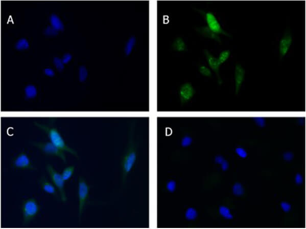
Immunofluorescence of Rabbit Anti-Beclin 1 Antibody. Cells: HeLa cells. Fixative: 100% MeOH. Permeabilizaton: 0.3% Triton X-100. Primary Antibody: Anti-Beclin-1 at 5ug/mL overnight at 2-8C. Secondary Antibody: Donkey Anti-Rabbit IgG DyLightTM488 (p/n 611-741-127) at 5ug/mL for 60mins at RT. Nuclear Counterstain: DAPI. Staining: (A) DAPI, (B) Anti-Beclin1+DyLight, (C) Merge A+B, (D) secondary only. Expected localization:: Golgi apparatus, mitochondria, endosome, nucleus.

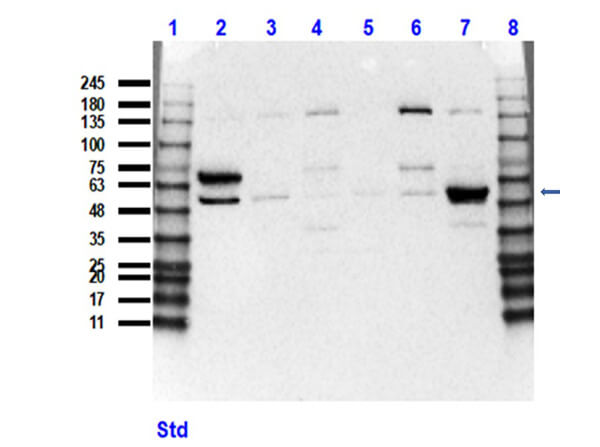
Western Blot of Rabbit Anti-Beclin 1 Antibody. Lane 1: Opal Prestain Molecular Weight Marker (p/n MB-210-0500). Lane 2: Beclin overexpressing HEK293T lysate (10ug) [+]. Lane 3: HEK293T vector vector (10ug) [-] (p/n W09-001-GX5). Lane 4: U87-MG whole cell lysate (35ug) [+]. Lane 5: C2C12 whole cell lysate (35ug) [+] (p/n W10-001-GL7). Lane 6: MOLT4 whole cell lysate (35ug) [+] (p/n W09-001-GL2). Lane 7: U251-MG whole cell lysate (35ug) [+] (p/n W09-001-GY4). Lane 8: Opal Prestain Molecular Weight Marker (p/n MB-210-0500). Primary Antibody: Anti-Beclin1 at 1:1000 overnight at 2-8C. Secondary Antibody: Goat Anti-Rabbit IgG HRP (p/n 611-103-122) at 1:40,000 for 60min at RT. Block: 5% BLOTTO/TBST (p/n B501-0500). Expected MW: ~51kDA endogenous, ~50-60kDa overexpressing. Observed MW: ~53, 75kDa.

|

|
|
Immunohistochemistry of Rabbit Anti Beclin 1 Antibody. Tissue: Human colon cancer tissue (40X). Fixative: none. Antigen Retrieval: Heat induced epitope retrieval (HIER). Primary Antibody: Anti Beclin 1 at 1:100 for 30mins at RT. Secondary Antibody: Anti Rabbit Poly HRP IgG ready to use 8 min at RT. Counterstain: hematoxylin. Expected Localization: nuclear or cytoplasmic staining of normal cells. Less staining of cancer cells. Result Analysis: Passed. Epithelial layer staining, nuclear staining, cell cell junction staining.
|
|
|
| メーカー |
品番 |
包装 |
|
RKL
|
600-401-MG4S
|
25 UL
|
※表示価格について
| 当社在庫 |
なし
|
| 納期目安 |
約10日
|
| 保存温度 |
-20℃
|
|


